19 Large Airway Disorders
The most common disorders include the following:
There are two types of tracheal collapse: dorsoventral and lateral. Dorsoventral collapse is much more common, in which the dorsal tracheal membrane (which is often redundant) is drawn into the tracheal lumen with changes in intratracheal pressure with respiration. This type of collapse can occur in the cervical region, the intrathoracic region, and can extend to involve the mainstem bronchi. Cervical collapse is associated more with inspiration because of the resultant drop in intratracheal pressure. Intrathoracic collapse is more associated with expiration. This results from intrathoracic pressure exceeding airway opening pressure. Dorsoventral collapse can be graded on a scale of 1 to 4, as described in Table 19-1. The grade is assigned to the most severely affected portion. Lateral collapse of the trachea is uncommonly diagnosed. The few reported cases have been seen in association with other factors: a congenital malformation, secondary to tracheal surgery, or possibly after trauma.
Table 19-1 Grades of Tracheal Collapse
| GRADE | TRACHEAL RING SHAPE | AIRWAY DIAMETER REDUCTION |
|---|---|---|
| I | Circular | 25% |
| II | Partially flattened | 50% |
| III | Nearly flat | 75% |
| IV | Flat to dorsally inverted | ≥90% |
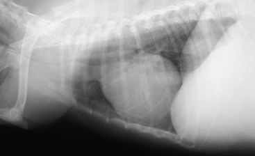
Thoracic auscultation may be difficult because of concurrent upper airway noise, increased bronchial sounds, and obesity. Concurrent cardiac disease may be present and must be ruled out as a cause for coughing. Systolic murmurs, pulmonary edema, or cardiac arrhythmias may be detected. Hepatomegaly is commonly detected, in most cases from fat deposition; however, it may also be a sign of cardiac failure. Heartworm status should be ascertained. Thoracic radiographs are the initial imaging study performed. Both inspiratory and expiratory films of the entire tracheal length should be obtained. This may demonstrate dynamic tracheal collapse (84% of cases in one study). Radiographs may be difficult to interpret with an overlying esophagus, positioning, and motion. Radiographs should be carefully evaluated for signs of cardiac disease, pneumonia, or other airway disease. Lateral thoracic radiographs (Fig. 19-1) taken at the level of the thoracic inlet commonly demonstrate tracheal collapse in dogs with severe coughing on expiration. In cases where collapsing trachea is suspected but not seen on thoracic radiographs, other diagnostics may be employed. Fluoroscopy shows dynamic tracheal movement and is better able to demonstrate collapse of mainstem bronchi, which is difficult to appreciate on survey radiographs. In some cases, stimulating the animal to cough will help localize the area of collapse. Ultrasonographic techniques for evaluating tracheal collapse have also been described, though these may be difficult to perform because of the air interface within the trachea. The gold standard for diagnosing collapsing trachea is tracheobronchoscopy. This technique allows evaluation of the entire trachea and the mainstem bronchi. Deeper airways may also be evaluated, depending on the size of the dog. The degree of tracheal collapse is also most accurately determined with an endoscopic procedure. Samples may be obtained from the airways for cytologic evaluation and microbial or fungal culture. This is important to rule out concurrent airway disease or possible infectious processes. Owners should be warned that there is the potential for worsening of respiratory signs after this procedure.

Figure 19-1 Lateral thoracic radiograph demonstrating significant tracheal collapse at the level of the thoracic inlet.
(Radiograph courtesy of Dr. E. Riedesel, Iowa State University.)
Medical and environmental management are the initial treatments for this condition. Environmental management includes minimizing excitement; using a harness instead of a neck collar; avoiding dust and smoke in the air by cleaning or removing carpeting, using air purifiers, etc.; and minimizing time spent in hot and humid environments to reduce panting.
Medical management must include weight loss for an obese animal. There may be concurrent allergic airway disease (diagnosed via radiography and bronchoalveolar lavage cytology) that may be treated medically. Concurrent respiratory infections may be observed with tracheal collapse; however, the true incidence of this is unknown. There is not sufficient evidence to warrant empiric antibiotic therapy in dogs with tracheal collapse. Antitussive agents are employed in therapy for both acute and chronic cases. Narcotic agents are the most potent cough suppressants, but may be associated with sedation, constipation, and loss of appetite. The use of bronchodilators is controversial. These agents do not act on large airways. However, they may be effective at lowering intrathoracic pressure and improving small airway obstruction. Side effects of these medications include cardiovascular stimulation, tachycardia, arrhythmias, and central nervous system stimulation. Corticosteroids may be used on a short-term basis, primarily for laryngeal or tracheal inflammation or for chronic airway disease. The potential side effects (weight gain, sodium retention, panting) should be measured against the possible benefits. All medications should be tapered to the lowest effective dose to minimize side effects and possible drug interactions. The common medications used for the medical treatment of tracheal collapse are listed in Table 19-2. Recently, there was a report on the use of diphenoxylate/atropine (Lomotil) for collapsing trachea. Diphenoxylate has a narcotic, antitussive effect. Atropine has two effects: reduction in mucus secretion and bronchodilation. There have been no published studies documenting the efficacy of this therapy. Surgical options are available for animals with severe disease or animals that are unresponsive to medical management. These options include both intrathoracic and extrathoracic stenting procedures.


