CHAPTER 58 Laboratory Diagnosis of Parasitic Diseases
Parasites that are covered in this chapter include helminths and protozoans. Some parasites are easily diagnosed because the diagnostic stages passed in the feces are readily detected and identified. Other parasites require more effort to identify because the eggs are not distinguishable; larval fecal cultures are needed to determine the species or groups of nematodes present, and some require special procedures to enhance diagnosis. This chapter assists the equine clinician in sample selection and interpretation of results to facilitate rapid and accurate diagnosis of parasitic diseases of horses.
HELMINTH DIAGNOSIS
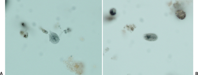
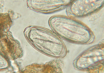
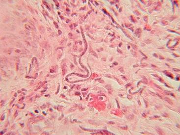
A large variety of nematode parasites infect horses. Most reside in the gastrointestinal tract and are detected by fecal examination; examples include the strongyles (Fig. 58-1), ascarids (Fig. 58-2), threadworms (Fig. 58-3), pinworms (Fig. 58-4), lungworms, and stomach worms. Some nematodes reside in solid tissues and do not pass any stages in the feces; these include the filarial worms, Onchocerca cervicalis, O. reticulata, and Parafilaria multipapillosa, which are detected by discovery of microfilariae, which are motile embryos (Fig. 58-5). Some worms cause problems as immature larval stages and do not produce any stages that leave the host. These are usually found by biopsy or are presumptive diagnoses, including summer sores caused by larval stomach worms, species of Draschia, and Habronema.
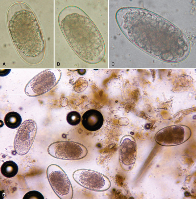
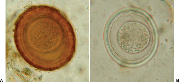
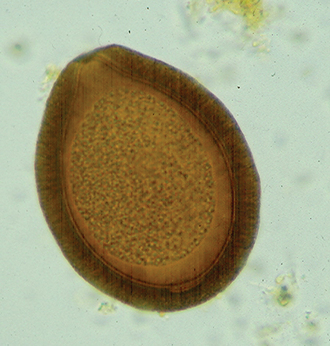
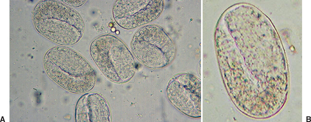
Fig. 58-2 A, Parascaris equorum egg (88 × 77 μm). B, P. equorum infertile and atypical egg (80 × 66 μm).
PROTOZOAN DIAGNOSIS
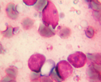
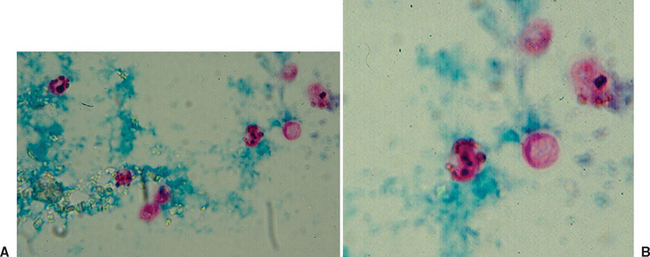
Very few protozoal organisms infect the gastrointestinal tract of horses, and some are believed to be beneficial as the ciliate fauna of the large bowel. Others are pathogenic or potentially pathogenic, including flagellates such as Giardia (Fig. 58-6) and possibly Leishmania (Fig. 58-7) and coccidians such as Eimeria (Fig. 58-8) and Cryptosporidium (Fig. 58-9).