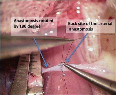
Fig. 8.1
Dissection of suprarenal vein
4.
Release both renal vessel from retroperitoneal tissue (Fig. 8.2)
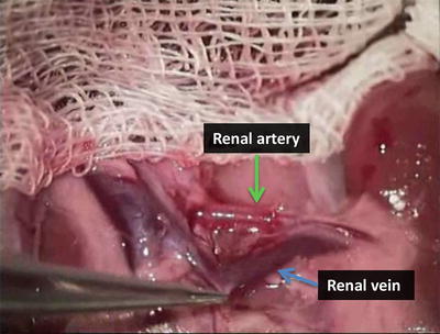

Fig. 8.2
Dissection of renal artery and vein
5.
Release the ureter from retroperitoneal tissue and cut them at the distal end (Fig. 8.3)
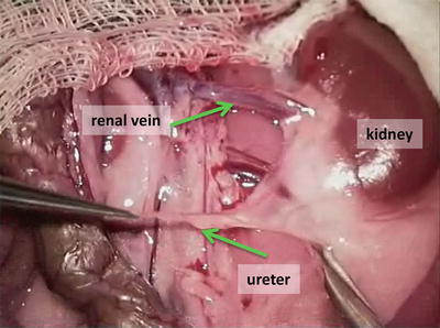

Fig. 8.3
Dissection of ureter
6.
Release the kidney from the surrounding tissue (Fig. 8.4)
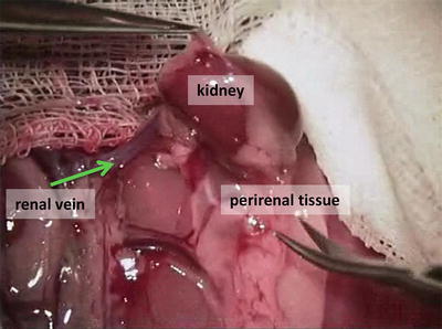

Fig. 8.4
Mobilization of the kidney from surrounding tissues
7.
Inject heparin solution (100 IU/1 ml) to the inferior caval vein
8.
Put the clamp on the aorta above the stump of the renal artery
9.
Cut the renal vessels as close as possible to the aorta and the caudal caval vein
10.
At the back table, remove the redundant tissue from distal part of the ureter and renal vessels so that the ends are prepared for anastomosis and perfuse the kidney with saline solution through renal artery (Fig. 8.5)
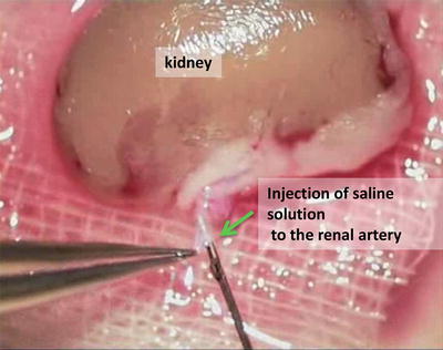

Fig. 8.5
Perfusion of the kidney
8.3 Kidney Graft Transplantation
8.3.1 Equipment and Materials
Scissors, forceps, four vascular clamps, 9-0 nylon sutures, 7-0 and 4-0 silk suture, two micro-scissors (straight, curved), three micro-forceps (two straight, one curved), micro-needle holder, thermal cauterize unit, cotton swabs, gauze, heating pad, needles, syringes (5 ml, 10 ml).
Protocol
1.
Open abdominal cavity with a midline incision
2.
Cauterize capillary bleeding from the incision edge
3.
Perform the recipient left nephrectomy so that the renal vessels and ureter are ligated and dissected as close as possible to the kidney hilus.
4.
Flush the renal vessels stumps with cold heparin saline solution (100 UI/1 ml saline)
5.
Start the arterial anastomosis with two fixating stitches (10/0) at the distance of 40 % in diameter from each other on the recipient renal artery (Fig. 8.6)
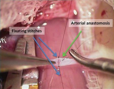

Fig. 8.6
Fixating polar stiches in arterial anastomosis
6.
Put four stitches equidistantly on the anterior site of the arterial anastomosis (Fig. 8.7)
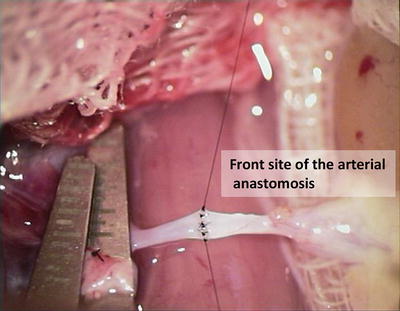

Fig. 8.7
Anterior wall of arterial anastomoses
7.
Rotate the artery by 180° to finish the posterior site of the anastomosis
8.
Put the five stitches equidistantly on the posterior site of the anastomosis (Fig. 8.8)

