Introduction to the Nematodes
Learning Objectives
After studying this chapter, the reader should be able to do the following:
• Briefly discuss the importance of nematodes in veterinary practice.
• Describe the important external morphologic features for identifying nematodes.
• Name the important parts of the male nematode’s reproductive system.
• Name the important parts of the female nematode’s reproductive system.
• Describe the four types of nematode eggs.
• Determine the difference among these reproductive terms: oviparous, ovoviviparous, and larviparous.
• Describe the “typical life cycle” of a representative nematode.
• Explain the difference between a direct life cycle and an indirect life cycle.
• Be able to integrate these terms with the nematode parasites covered in Chapter 4.
Key Terms
Cervical alae
Copulatory bursa
Bursal rays
Spicules
Dioecious
Ascaroid- or ascarid-type egg
Trichostrongyle, strongyle-type egg
Spiruroid-type egg
Trichinelloid-trichuroid-type egg
Oviparous nematode
Ovoviviparous nematode
Larviparous nematode
Molt
Infective third-stage larva
Direct life cycle
Indirect life cycle
Members of the phylum Nematoda, the nematodes, or roundworms, are the most numerous and most diverse group of animals on Earth. Approximately 10,000 species thrive in very diverse habitats. There are three basic types of nematodes: (1) the free-living nematodes residing in marine water, freshwater, and soil environments; (2) the nematodes that parasitize plants; and (3) the nematodes that parasitize domesticated and wild animals and humans. This chapter discusses the third category, nematodes that parasitize domesticated and wild animals and, to some extent, humans.
 TECHNICIAN’S NOTE
Nematodes are collectively referred to as roundworms and are the most numerous and diverse group of animals on Earth.
Nematodes of Importance in Veterinary Medicine
Nematodes in domesticated farm animals in the United States alone cause losses of billions of dollars in animal agriculture resulting from veterinary bills and death. Plant nematodes cause approximately 10% of cultivated crops to be lost each year. It is impossible to determine similar monetary losses in the pet or companion animal “industry.”
Key Morphologic Features
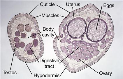
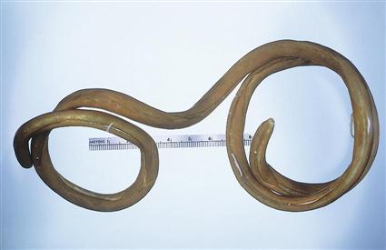
Protozoans, discussed in Chapters 10 and 11, are unicellular organisms; nematodes, however, are multicellular organisms. Whereas trematodes (leaflike in form) and cestodes (chainlike or boxcarlike in form) are dorsoventrally flattened in cross section, nematodes are unsegmented, elongate, rounded on both ends, circular in cross section, and bilaterally symmetric (Figure 3-1); thus they are often referred to as roundworms. Nematodes come in a variety of sizes, from Strongyloides stercoralis, a tiny parasitic female worm only 2 mm in length and about 35 µm wide (Figure 3-2), to Dioctophyma renale, “the giant kidney worm,” measuring 100 cm (1 m) in length and 1.2 cm wide (Figure 3-3). As mentioned previously, most nematodes are cylindric and rounded on both ends; however, nematodes come in a wide variety of shapes, from spherical (definitely not cylindric), such as Tetrameres species, parasites of the gizzard of domesticated and wild birds (Figure 3-4), to the whiplike Trichuris species, which parasitize every domesticated animal except the horse (Figure 3-5).
 TECHNICIAN’S NOTE
Nematodes are referred to as roundworms because they are circular in cross section, rounded at both ends, unsegmented, elongate, and bilaterally symmetric.
External Morphologic Features
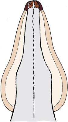
Nematodes are covered by a thin cuticle (see Figure 3-1). This cuticle covers the exterior body surface of the nematode and extends into all its body openings, that is, the mouth, esophagus, and rectal and genital openings. On the external surface the cuticle has a variety of modifications. There may be lateral flattened expansions of the cuticle in the region of the anterior end; these expansions are called cervical alae (Figure 3-6). There also may be lateral flattened expansions of the cuticle in the region of the posterior end of some male nematodes; this posterior lateral expansion is called the copulatory bursa and serves to hold onto or to grasp the female nematode during the mating process (Figure 3-7). The copulatory bursa is composed of fingerlike projections called bursal rays. Between each adjacent bursal ray are thin membranes. The male’s highly chitinous and pigmented spicules (see Figure 3-7) (the intromittent organs, or “penis”) are associated with the copulatory bursa. The cervical alae and the copulatory bursa are only two of the many modifications of the external cuticle of nematodes. The nematode’s cuticle is secreted, or formed, by the thin layer directly beneath it, the hypodermis (see Figure 3-1).
Stay updated, free articles. Join our Telegram channel

Full access? Get Clinical Tree


