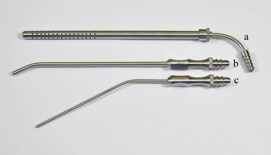
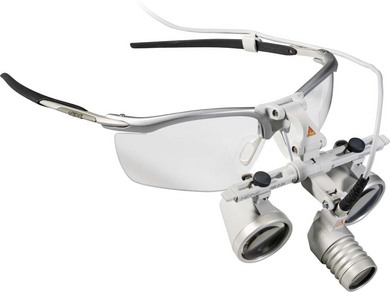
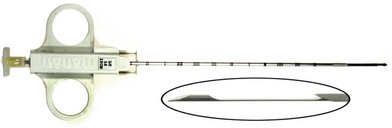
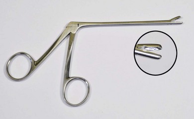
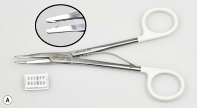
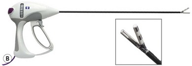
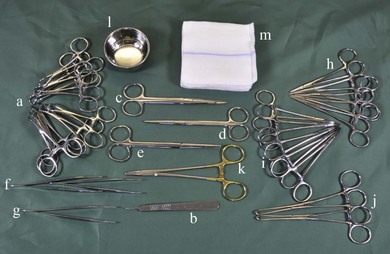
Chapter 13 General surgical instruments including scalpels, dissecting forceps, scissors, artery forceps, retractors, and needleholders are described in the chapter on Instrumentation in Feline Orthopedic Surgery and Musculoskeletal Disease (Chapter 23).1 An example of the instruments that could be included in a general surgical kit is listed in Box 13-1 and shown in Figure 13-1. Figure 13-1 Surgical tray. a, Backhouse towel clips 9 cm; b, number 3 scalpel handle; c, Mayo scissors straight 14 cm; d, Metzenbaum scissors straight 14.5 cm; e, Metzenbaum scissors curved 14.5 cm; f, Adson Brown dissecting forceps; g, Adson Debakey dissecting forceps; h, Halstead mosquito artery forceps straight 12.5 cm; i, Halstead mosquito artery forceps curved 12.5 cm; j, Allis tissue forceps 4/5 teeth 15 cm; k, Mayo-Hegar (T.C.) needleholder 15 cm; l, gallipot; m, sponges with radio dense marker. Suction will contribute to a clear operating field. Many tips are available but the most useful focal tips are the Adson (4 mm diameter) or Frazier (2.6 mm diameter) types, both of which offer fingertip control of suction by an assistant. For suction of larger volumes of fluid from body cavities when performing abdominal or thoracic surgery the Poole is preferred (Fig. 13-2). Use of binocular loupes, which magnify while maintaining a normal working distance, will significantly improve visualization of the operative site. A magnification of 2.5× is sufficient for most procedures. The focal distance should be selected to maintain a normal working distance, usually 380 mm or 430 mm. The addition of a loupe light, either fiber optic or the more economic LED type, will automatically provide surgery light where it is needed (Fig. 13-3). Selection of the appropriate soft tissue biopsy devices will be detailed in the relevant organ specific chapters and more generally in Chapter 14. Several types are available. The TruCut type consists of a slim needle which may be inserted relatively atraumatically into an area of parenchymatous tissue, typically within an organ (Fig. 13-4). A spring loaded cutter is fired within the needle, which excises and traps a sample. Where the sample is to be obtained from the surface of a friable tissue, clamshell type biopsy forceps with cutting jaws enclose and remove the sample (Fig. 13-5). Skin biopsies may be obtained using a punch biopsy, available in a variety of sizes. Although available as a reusable instrument it is normally used as a disposable device. The following were compared for their efficacy and security in achieving vessel ligation: braided suture in a surgeon’s knot; a monofilament suture in a granny knot; a metallic clip (Ligaclip, Johnson and Johnson; Fig. 13-6A); a bipolar diathermy system (Ligasure, ValleyLab; Fig. 13-6B); and an ultrasonically activated scalpel (Harmonic Scalpel, Johnson and Johnson).2 Secure hemostasis was obtained with all the techniques in all vessels below 5 mm in diameter. In vessels over 5 mm, secure hemostasis was obtained with all modalities except the harmonic scalpel. With the harmonic scalpel, leaks occurred in vessels greater than 5–6 mm, confirming that the manufacturer’s claims for each of the hemostatic methods were accurate. The use of hemostatic tools such as the harmonic scalpel and Ligasure has increased with the use of laparoscopic surgery where it is not easy to place traditional surgical knots.
Instruments
General surgical instruments
Suction
Magnification and light
Soft tissue biopsy instruments
Vessel sealing devices
![]()
Stay updated, free articles. Join our Telegram channel

Full access? Get Clinical Tree


Instruments