Imaging of Myelin by Coherent Anti-Stokes Raman Scattering Microscopy
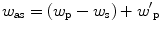
) frequencies interact with molecules in a medium and generate a new field at the anti-Stokes frequency . In most experiments, the pump field Ep and probe field come from the same laser beam. When the beating frequency (ωp−ωs) is in resonance with a vibration of the molecular chemical bonds, the mixed pump and Stokes field can effectively excite the molecule to a vibrationally excited state. The efficiency of generated CARS process is then significantly enhanced because the molecular vibration facilitates the energy exchange between light fields. Therefore, the molecules in resonance will show higher signal than those of resonance, providing a vibrational contrast in a CARS image. Meanwhile, as a coherent process, the CARS signal fields from different molecules have a well-defined phase relationship and they add up like the emission from an antenna array. The emission is concentrated in one direction defined by the constructive interference, which greatly facilitates the signal collection. The coherent addition also results in a quadratic signal increase with the respect to the density of molecular chemical bonds.
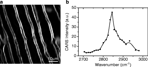
In the nervous system, myelin sheath is a multiple layer membrane which comprises 70% lipid by weight. Membrane lipid contains a huge amount of ordered CH2 groups in the lipid chains. The vibrations of these ordered CH2 groups produce strong CARS signal. Figure 1a shows a CARS image of myelin sheath in the fresh spinal tissue isolated from guinea pig and a CARS spectrum of myelin (12). The strong peak in the CARS spectrum represents the symmetric stretching vibration at 2,840 cm−1 (Fig. 1b). CARS microscopy offers several unique advantages for myelin imaging.
Fig. 1.
CARS image and CARS spectrum. (a) A typical CARS image of myelin sheath in the fresh spinal tissue isolated from guinea pig. (b) CARS spectrum of myelin sheath showing a strong peak at 2,840 cm−1, which represents symmetrical stretching vibration of CH2 groups.
1.
Label-free detection of myelin
CARS microscopy directly probes the symmetrical stretching vibrations of CH2 groups in the myelin lipids, thus no label is necessary for CARS imaging of myelin. Nonlabeling property simplifies the sample preparation and avoids the photobleaching problem occurring at the imaging using fluorescent labels. Therefore, CARS imaging is nondestructive and can be applied to image myelin in its natural state, such as in biologically active tissues and live animals (13).
2.
3D submicron resolution
The nonlinear dependence on excitation intensity ensures that the CARS signal is only generated in the focal center, providing an inherent submicron 3D spatial resolution. For myelin imaging, the lateral resolution was measured to be 0.28 μm and the axial resolution was calculated to be 0.70 μm (12). Such resolution allows detecting the detailed structure in myelin, such as a node of Ranvier.
3.
Multimodal nonlinear optical imaging
The laser beams for CARS could be used for two-photon excited fluorescence (TPEF) and sum-frequency generation (SFG)/second harmonic generation (SHG) so that TPEF and SFG/SHG imaging can be performed simultaneously with CARS imaging in the same platform. In this multimodal nonlinear optical imaging platform, CARS is used for imaging of myelin; TPEF is used for imaging of fluorescent probe-labeled ions or proteins, such as Ca2+ ion indicators; SFG/SHG is used for imaging of astroglial filaments (14). Therefore, multiple components in the nervous tissues can be visualized at the same time.
4.
Raman spectral information of myelin lipid
In addition to morphology of myelin, conformation of the hydrocarbon chains of myelin lipid is also able to be determined by confocal Raman spectroscopy in the CARS platform (15). Raman spectra of myelin lipid from the C–C and C–H vibrational bands can be used to determine the intermolecular chain disordering degree of myelin lipid and Raman spectra from C = C bonds and C–H bonds in lipid acyl chains can be used to determine the unsaturation degree of myelin lipid chains. The information obtained from Raman spectra will help to analyze the change of myelin lipid in the diseased condition.
3 CARS Microscope
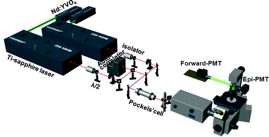
Figure 2 shows a schematic of a CARS microscope which includes a laser source, optical pathway, a laser-scanning microscope, and signal collection components. The details of these four parts are described below.
Fig. 2.
Schematic of a CARS imaging setup.
1.
A laser source: The production of a CARS process requires a pump and a Stokes laser beam. In Fig. 2, both lasers are generated from two Ti:sapphire oscillators (Mira 900, Coherent Inc, Santa Clara, CA) and they are tunable from 700 nm to 1,000 nm, where water absorption is minimized. The pulse width is 2.5 ps whose corresponding spectral width matches the line width of Raman bands. The laser source for CARS can also be realized by using optical parametric oscillator (OPO) pumped by a pulsed laser (16).
2.
Optical pathway: In Fig. 2, collinear beam geometry is utilized to fulfill the phase matching condition which also simplifies the implementation. The two laser beams are tightly synchronized (Sync-Lock, Coherent Inc) with an average timing jitter of 100 fs. The two beams are collinearly combined using a dichroic combiner (LWP-45-R720-7850-PW-1004-UV, CVI Laser LLC, Albuquerque, NM). A Pockels cell (Model 350-160, Conoptics, Danbury, CT) is used to lower the repetition rate, which reduces average power but maintains high peak power at the sample. The pockels cell is important in the imaging of fixed slices where a low laser power is needed to minimize the photodamage. In the imaging of fresh tissues and live animals where higher laser power is required, the pockels cell can be removed from the optical pathway. The polarization of the incident beams is controlled by half-wave plates.
3.
Laser-scanning microscope: Two collinearly combined beams are directed into a laser scanning confocal microscope (FV300/IX70, Olympus America Inc, Melville, New York). To produce a high-quality CARS image, a high numerical aperture (NA) objective, such as a 60× water objective (NA = 1.2) or a 40× water objective (NA = 0.8) is used to form tightly focused excitation beams through the sample. To obtain a CARS image in a large field of view, a 20× air objective (NA = 0.75) is also acceptable to generate CARS signal.
4.
Signal collection: The forward-detected CARS (F-CARS) signal is collected by an air condenser (NA = 0.55). The epi-detected CARS (E-CARS) signal is collected by the same objective. In multimodal nonlinear optical imaging, the epi-detected TPEF or SFG/SHG signal is spectrally separated from the E-CARS signal by a dichroic splitter. CARS signal from relatively thin fixed slices or cryosections is favorably collected in forward direction. CARS signal from fresh tissues is able to be collected in both forward and epi-direction. CARS signal from live animals can only be collected in epi-direction. Both F-CARS and E-CARS signals are detected with the same type of photomultiplier tube (PMT, R3896, Hamamatsu, Japan). On the CARS microscope, a spectrometer equipped with an electron multiplying charge coupled device (EMCCD) can also be attached to the side port with the slit replaced with a pinhole for confocal Raman analysis.
4 Polarization Dependence of CARS Signal
CARS signal is sensitive to the polarization of excitation beams. CARS signal from myelin (III) is maximized when the polarization of two excitation beams is parallel to the symmetrical axis of CH2 groups in myelin lipid and CARS signal (I┴⊥) is minimized when the polarization is perpendicular to the symmetrical axis of CH2 groups. Therefore, in a CARS microscope, half way plates are installed to adjust the polarization of excitation beams according to the orientation of myelin fibers in the sample.
The polarization dependence of CARS signal is able to be used to determine the orientation and ordering degree of lipid in myelin based on the ratio of CARS signals (III/I┴⊥). The information of ordering degree of lipid in myelin can be used to determine the myelin condition during the degradation process (17). On the other hand, the influence of polarization dependence of CARS signal is able to be eliminated through reconstruction of the two complementary images with perpendicular excitation polarizations (18) or by using circularly polarized laser beams (19).
5 Ex Vivo CARS Imaging of Spinal and Brain Tissues
Materials used for CARS imaging of myelin in fresh, fixed, and frozen tissues:
(a)
Krebs’ solution: NaCl 124 mM, KCl 2 mM, KH2PO4 1.2 mM, MgSO4 1.3 mM, CaCl2 2 mM, dextrose 10 mM, NaHCO3 26 mM, and sodium ascorbate 10 mM, equilibrated with 95% O2, 5% CO2; or other artificial cerebrospinal fluid;
 . In most experiments, the pump field E p and probe field
  come from the same laser beam. When the beating frequency (ω p−ω s) is in resonance with a vibration of the molecular chemical bonds, the mixed pump and Stokes field can effectively excite the molecule to a vibrationally excited state. The efficiency of generated CARS process is then significantly enhanced because the molecular vibration facilitates the energy exchange between light fields. Therefore, the molecules in resonance will show higher signal than those of resonance, providing a vibrational contrast in a CARS image. Meanwhile, as a coherent process, the CARS signal fields from different molecules have a well-defined phase relationship and they add up like the emission from an antenna array. The emission is concentrated in one direction defined by the constructive interference, which greatly facilitates the signal collection. The coherent addition also results in a quadratic signal increase with the respect to the density of molecular chemical bonds.