GLUCOSE

34 GLUCOSE


1 Why does delayed processing or analysis of a blood sample result in a lower glucose concentration?


Prolonged exposure of serum or plasma to leukocytes, platelets, and erythrocytes allows the cells to consume glucose and lower the glucose concentration. The blood cells use the plasma glucose for their glycolytic pathways. The decline of plasma or serum glucose concentration is generally 5% to 10% per hour. This usually does not interfere with the interpretation of glucose concentrations because the processing of patient samples and the samples used for reference interval determination should be the same (i.e., removing serum or plasma from cells within 30 to 60 minutes after blood collection). The rate of glucose consumption can increase when there are more cells in the blood (marked leukocytosis, marked thrombocytosis, or possibly erythrocytosis). The rate of glucose consumption can be decreased by placing the blood sample in a cool environment, but clot formation and contraction are also reduced.


Serum separator tubes (tubes with gold or red/black stoppers) contain an activator and gel that enhance clot formation and allow easier separation of a blood clot and serum, respectively. After centrifugation, the serum glucose concentration remains stable in a refrigerator for at least 48 hours if the gel barrier is intact.


Erythrocytes from pigs are unique in that they lack a functional glucose transporter and therefore utilize glucose at a much lower rate than erythrocytes from other species. Inosine is the major energy substrate in pig erythrocytes, whereas glucose is the primary substrate in other domestic species.


10 What are the three major changes or alterations in physiologic processes that produce a hyperglycemia?


Hyperglycemia can be created by (a) increased glucose intake, (b) increased glucose production, and/or (c) decreased glucose uptake by peripheral tissues.


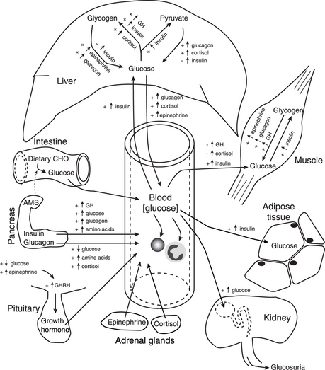
Increased glucose intake can result from ingestion of carbohydrate meal or the infusion of a glucose-containing fluid (e.g., 5% dextrose). Strictly speaking, increased glucose production is limited to increased gluconeogenesis within hepatocytes. However, hepatic glycogenolysis can also result in the release of glucose into blood. Decreased glucose uptake by peripheral tissues can result in a mild hyperglycemia by itself, but increased glucose uptake or increased glucose production can enhance the severity of the hyperglycemia. Figure 34-1 illustrates how these processes are influenced by activities of hormones and other factors.

image

Figure 34-1 Physiologic factors that influence blood glucose concentration.


Intestine: In monogastric animals, dietary carbohydrates (CHO) are broken down to monosaccharides (including glucose) that are absorbed in the small intestine, from which they enter portal blood and then systemic blood if not removed by hepatocytes.


Pancreas: Insulin and glucagon are released from pancreatic islet cells (beta cells and alpha cells, respectively). Insulin secretion is stimulated by increased blood concentrations of glucose, growth hormone (GH), glucagon, and amino acids. Glucagon secretion is stimulated by increased blood concentrations of amino acids and cortisol or by decreased blood glucose concentrations.


Liver: Hepatocytes are the primary source of blood glucose during fasting. Glucose can be obtained from glycogenolysis (stimulated by epinephrine and glucagon but inhibited by insulin) or gluconeogenesis (stimulated by glucagon and cortisol but inhibited by insulin). Insulin also promotes glycolysis. Increased glucose release from hepatocytes is promoted by increased glucagon, cortisol, or epinephrine. Insulin promotes the uptake of glucose by promoting glucokinase activity.


Muscle: Glucose uptake by myocytes is promoted by insulin through specific insulin receptors and glucose transporters; GH and cortisol inhibit the uptake of glucose. Insulin promotes glycogen synthesis in myocytes, whereas GH, glucagon (in cardiac muscle), and epinephrine promote glycogenolysis.


Adipose tissue: Insulin promotes the uptake of glucose by adipocytes through specific insulin receptors and glucose transporters.


Kidney: If the renal threshold is exceeded, hyperglycemic glucosuria will develop.


Pituitary: GH release from the pituitary is stimulated by growth hormone–releasing hormone (GHRH), which is released from the hypothalamus during hypoglycemia or after epinephrine stimulation.


Blood cells: Glucose enters erythrocytes (except porcine), leukocytes, and platelets through insulin-independent processes and is used in glycolysis and the hexose monophosphate shunt. Pig erythrocytes lack a functional glucose transporter and use inosine as the major energy substrate.


(Modified from Stockham SL, Scott MA: Fundamentals of veterinary clinical pathology, Ames 2002, Iowa State Press.


Aug 26, 2016 | Posted by in INTERNAL MEDICINE | Comments Off on GLUCOSE

Full access? Get Clinical Tree

Get Clinical Tree app for offline access