35 GASTROINTESTINAL TRACT
6 Which basic tests should be performed in acute and chronic cases of diarrhea?
Tests should include fecal examination for parasites, hematology, and serum biochemistry.
7 Which gastrointestinal results can be expected on the complete blood count?
Red cell parameters
An increase in the packed cell volume (PCV) associated with increased total protein indicates hemoconcentration (dehydration). Conversely, a normocytic, normochromic, nonregenerative anemia may indicate chronic inflammation or, if the condition is acute, acute GI blood loss. This anemia of acute blood loss if frequently associated with a panhypoproteinemia. In cases of chronic GI blood loss (e.g., secondary to hookworms, ulcers, or tumors), an iron deficiency anemia may develop. Importantly, iron deficiency anemia secondary to blood loss may be regenerative, and therefore the mean corpuscular volume (MCV) may be within reference limits despite evidence of microcytosis on blood smears. Microscopic examination of a blood smear is always indicated in this case, since microcytosis will appear on a blood smear before the MCV begins to decrease.
8 Which gastrointestinal results can be expected on the biochemistry profile?
9 If small intestinal bleeding is suspected and the animal does not show obvious signs of melena, how reliable are the tests for occult blood in feces?
11 Describe the clinicopathologic changes associated with canine parvovirosis.
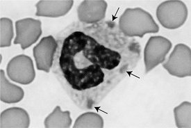
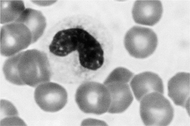
In addition to the clinical signs of acute hemorrhagic diarrhea, fever, and dehydration in young animals, dogs with parvovirosis are frequently leukopenic, neutropenic, and lymphopenic, with toxic neutrophils and a left shift (Figures 35-1 and 35-2). This hematologic change is present in up to 90% of infected animal. A leukopenia of less than 1500 to 2000 white blood cells per microliter (WBCs/μl) is common. The neutropenia is secondary to both increased losses of neutrophils in the gut and impaired bone marrow production of neutrophils, because the parvovirus is toxic to rapidly dividing bone marrow cells. Toxic neutrophils and left shift are secondary to the increased demand of neutrophils in the GI tract. Note that some breeds are more susceptible to parvovirus and include Rottweilers, Dobermans, pit bulls, and English Springer spaniels.

Figure 35-1 Toxic neutrophil in blood of a cat with panleukopenia. Note the presence of several Döhle bodies (arrows).
(Modified Wright’s stain; 500×.)
16 What hematologic changes can be expected in canine salmonellosis?
Hematologic changes may be nonspecific, but frequently a severe leukopenia or, conversely, a neutrophilia can be present. In almost all cases, however, a left shift with toxic neutrophils is present, reflecting an increased demand in peripheral tissues (see Figures 35-1 and 35-2).
17 How is salmonellosis confirmed?
Bacterial culture of feces is essential for the final diagnosis of salmonellosis.
21 Besides clinical signs, do clinicopathologic changes support a diagnosis of hemorrhagic gastroenteritis?
24 Describe the clinicopathologic changes in cases of feline panleukopenia?
As its name indicates, this virus causes a decrease in all WBC lines, primarily neutrophils and lymphocytes. A WBC count less than 2000/μl is common and frequently associated with a left shift and toxic neutrophils (see Figures 35-1 and 35-2).
Stay updated, free articles. Join our Telegram channel

Full access? Get Clinical Tree


