Fungal Infections

CHAPTER 17 Fungal Infections


Cutaneous Fungal Infections


Dermatophytes


The majority of clinical cases presented are caused by three fungi: Microsporum canis, Microsporum gypseum, and Trichophyton mentagrophytes. Typically M. canis is the most common source of dermatophytosis in dogs and cats. Table 17-1 summarizes dermatophytes isolated from hair and skin of dogs and cats.


TABLE 17-1 Dermatophytes isolated from dogs and cats


Most common
Microsporum Most common isolate
Microsporum gypseum Generally from soil contact
Trichophyton mentagrophytes Soil contact


Less common
Microsporum persicolor Sylvatic form
Microsporum fulvum Rare
Microsporum audouinii Rare
Trichophyton rubrum Rare

The dermatophytic fungi are spread through contact with infected hair or scales or contaminated environments or through carriage by fomites. Infective spores enter the environment when contaminated hairs break and are shed. Combs, brushes, clippers, bedding, transport cases, and paraphernalia associated with grooming, moving, or housing of animals can be sources of infection and subsequent reinfection. These spores are small and can be transported by currents of air, on dust, and on other mechanical fomites.


M. canis infections are usually caused by exposure to an infected cat or contact with fomites contaminated by an infected cat. The incubation period is from 1 to 3 weeks. Germination is temperature dependent. Most Trichophyton infections are caused through contact with infected rodents, their nests, or contaminated fomites. Dogs and cats are infected by the geophilic fungal organism M. gypseum from rooting, sleeping, and digging in contaminated soil. Animals can be exposed by reverse zoonoses from direct contact with infected people.


Dermatophytic infections display spontaneous resolution when infected hairs either enter the telogen phase or an inflammatory reaction is triggered. When hair follicles reach telogen, the production of keratin slows and stops. Dermatophyte infections require actively growing hair to survive. Infective arthrospores remain on the hair shaft, but reinfection of that particular hair follicle does not reoccur until that follicle reenters the anagen phase. Dermatophyte infections are often self-limiting and resolve spontaneously within 3 to 4 months.


Younger animals are predisposed to acquiring and developing symptomatic dermatophyte infections. This is in part because of a delay in development of adequate host immunity and immature immune surveillance machinery. However, there are also differences in puppies and kittens in biochemical properties of the hair and skin (especially sebum), the growth and replacement of hair, in healing properties, and in the physiologic status of the host as related to age that may also play a large role.


Clinical Signs


Dermatophytosis is a diagnosis that must be based on positive culture results or biopsy. In multiple studies of suspected fungal skin cases, only about 15% were positive on culture. Results from all studies of cutaneous diseases of dogs and cats where fungal cultures have been obtained show the prevalence of dermatophytic infections is approximately only 2% of all dermatologic disease cases.


Fungal skin infection is almost always a disease of the follicles in dogs and cats. The most common clinical signs include hair loss, scaling, and crusting. There may be one or many circular patches with variable degrees of scaling. Some animals demonstrate the classic ring lesion with central healing, fine follicular papules, and crusts at the periphery. Pruritus is variable and is usually minimal or absent.


Dermatophytic lesions in puppies typically consist of focal or multifocal areas of hair loss and are clinically indistinguishable from other sources of hair loss. Dermatophytes invade hair shafts and cornified epithelium. As a result, the hair shaft is destroyed and normal keratinization is disrupted. Clinically this results in scaling and hair loss, but fungal skin infections can have a wide array of presentations, particularly if pruritus and secondary bacterial infection are involved. Dogs are more likely than cats to develop the classic ring lesion, a focus of alopecia, follicular papules, scales and crusts, and a central area of hyperpigmentation.


Infections with Trichophyton may also cause folliculitis and furunculosis of one paw or leg. Like the facial form, this leg or foot furuncular form can leave significant scars once healed. Generalized infections can present as a seborrhea-like eruption with greasy scales. The dermatophyte version is an exudative, usually fairly well circumscribed, nodular type of lesion that may develop multiple draining tracts. It is most often in dogs associated with M. gypseum or T. mentagrophytes infections. It can commonly appear as a solitary lesion on the face or on a limb. Although onychomycosis is rare, it is typically produced by T. mentagrophytes and presents asymmetrically with only one digit or multiple digits on only one paw involved.


In cats, dermatophytosis can effectively mimic almost any described feline skin disease. More than 90% of the time M. canis is the culprit responsible for feline dermatophytosis. M. canis is not typically a localized disease in cats.


In kittens, dermatophytes tend to consist of areas of alopecia and scaling. Erythema is variable and hard to detect in both long-haired and dark-haired cats. Lesions usually first appear as areas of hair loss on the face, muzzle, ears, and forelegs. Extent and severity of the lesion are inextricably tied to the health of the kitten. As a result, lesions can be focal, multifocal, or generalized. In some young cats, M. canis can produce comedone-like lesions resembling chin acne.


Diagnosis


Fungal culture


Fungal culture of affected material (scale and hairs) is the most reliable method to diagnose dermatophytosis and the only way to identify specific fungal pathogens. However, both false-positive and false-negative results are possible. Proper specimen collection techniques are critical for successful diagnosis.


Specimens are best collected by plucking hair near the lesion and active inflammation. Hair can be plucked with a hemostat or forceps or through use of a “brushing” method. This involves a brand-new (mycologically sterile) human toothbrush vigorously combed over suspect areas for 2 to 3 minutes. The bristles of the toothbrush (and their attached hairs) are lightly and repeatedly pressed into the surface of the culture medium.


In kittens, brushing both the face and the hair inside the bell of the ear is extremely important because early lesions often start at these sites. In the case of suspect claw or nail bed infection, special culture techniques may be necessary.


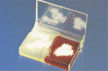
Dermatophyte test medium can be easily obtained for in-house procedures and is best bought as a plate for the toothbrush technique (Figure 17-2). Dermatophyte medium containers are best incubated loosely covered or capped at room temperature protected from ultraviolet light and desiccation, and covered in a container holding a damp paper towel. Colonies of dermatophytes can appear within 5 to 7 days of inoculation. Nevertheless, plates must be maintained for at least 3 weeks before determining a culture negative. Culture plates must be examined daily for medium color change to red. Growth of a white to buff-colored, powdery to cottony mycelium also will be observed. Color change must occur simultaneously as the first colony is visible, never later. All fungi, including nonpathogen fungal growth, will produce a red color change after colonies have growth from several days to a week. Colonies of infectious dermatophytes are never black, brown, gray, or green. Often M. canis will fail to grow or sporulate on dermatophyte medium. Recently studies have demonstrated that increased sporulation and growth can be obtained when plates were incubated at higher temperatures of 70° to 73° F (21° to 23.8° C).


Most colonies will produce spores after 7 to 10 days of growth, which will allow specific identification on microscopy. Spore collection is best accomplished by brushing clear cellophane tape lightly over the colony surface. Place the tape sticky side down onto a drop of lactophenol cotton blue stain on a glass slide. Then add another drop of stain on top of the tape. After a coverslip is placed, the prep can be examined at 100× magnification. Among the hypha strands will be microconidia (spores) that will have characteristic shapes according to their species. If no spores are visible, wait another 4 to 7 days. Certain colonies may not sporulate until they are old and established. The presence of an infectious dermatophyte can only be definitively confirmed by culture results, appearance of the colonies, and microscopic confirmation of fungal elements.


Treatment


Clinical management and therapeutic regimens for cutaneous fungal infections in puppies and kittens are complicated by the immature immune systems; hepatic, renal, and healing mechanisms of the young; and often the exceedingly small size of these animals.


Healthy dogs and some cats may show spontaneous remission of dermatophyte infections within 3 months. Kittens and cats with seemingly local disease can self-cure, but infections may be prolonged, at least 60 to 100 days. Long-haired cats can have spontaneous remission, but it may take from 1.5 to 4 years. Animals with generalized dermatophytosis typically require systemic therapy. For dogs, sylvatic forms of ringworm (Microsporum persicolor and Trichophyton species) do not resolve spontaneously and need systemic therapy.


Ideal clinical therapy involves not only treatment of the infected animal but also treatment of all animals exposed and the environment. Topical therapy used in conjunction with systemic treatment results in a faster mycologic cure than systemic therapy alone and reduces contamination on the haircoat and subsequent environmental contamination.


Topical treatment


Localized or spot treatments of dermatophyte infections in companion animals have almost no evidence for their efficacy. More effective than topical “spot” treatment are whole-body shampoos, rinses, or dipping with topical antifungal medications. The most effective topical antifungal whole-body baths, rinses, and dips are lime sulfur, enilconazole, and miconazole. Captan, povidone-iodine, and chlorhexidine are ineffective against dermatophytes when used as whole-body topical treatments.


Lime sulfur has shown superior antifungal activity at 8 oz per gallon of water (a 1:16 dilution). Lime sulfur applied twice weekly at 4 oz per gallon has demonstrated to be effective against dermatophytes if used together with whole-body clipping and aggressive environmental treatment methods. Lime sulfur is virtually nontoxic if applied properly and can even be safely applied to newborn puppies and kittens.


Enilconazole is effective in treating dermatophytosis when used as a sole, whole-body therapy (dipping) following whole-body hair clipping. Enilconazole is generally well tolerated. Side effects can include hypersalivation, anorexia, weight loss, and generalized muscle weakness. There have been anecdotal accounts of severe toxicity in cats after ingestion of enilconazole. It is believed these cases result from cats ingesting the medication by grooming after application of the antifungal agent. It appears that enilconazole is safe for cats if they are fitted with Elizabethan collars for a few hours after treatment and grooming is prevented until the animal is dry. Enilconazole is an effective treatment as a topical medication (10% solution or 100 mg/ml), but it is licensed only for dogs and horses.


Miconazole can be used as a sole therapeutic agent or used together in combination with chlorhexidine. Generally it is used twice weekly as an adjunct topical to systemic therapy rather than as a sole treatment method. As with all medicated shampoos and dips, for optimal therapeutic effects a skin contact time of 10 minutes is recommended for miconazole. Synergism between miconazole and chlorhexidine has been confirmed, and shampoos with this combination have been shown to hasten mycological cure. A list of topical antifungal therapy is included in Box 17-1.


Systemic treatment


Systemic therapy of dermatophytosis is used to hasten resolution of infection. Several drugs have been shown effective in the treatment of dermatophytosis. The appropriate choice for treatment is based on fungal species present, patient species involved, age and size of the patient involved, possible adverse effects and toxicity, and cost. Dogs and cats with multifocal lesions, all long-haired animals, and those in multianimal situations are candidates for systemic antifungal treatment. Animals nonresponsive following 2 to 4 weeks of topical therapy should also be considered for systemic treatment. See Box 17-2 for dermatophyte treatment summary. Itraconazole is currently the systemic treatment of choice for dermatophytosis in dogs and cats. Griseofulvin and ketoconazole both have significant issues of safety and efficacy. Box 17-3 and Table 17-2 list systemic fungal medications and antifungal drug dosages.


BOX 17-3 Systemic fungal medications


Amphotericin B is the only drug that is fungicidal and has been proven capable of permanently clearing CSF infections. Use of this drug requires hospitalization because it must be given parenterally and can be nephrotoxic in dogs or cats with disseminated infection. Newer forms of amphotericin (lipid complex and liposomal forms) are less nephrotoxic, although much more expensive. Pretreating with heat (at 140° to 158° F, 60° to 70° C for 10 minutes) reduces nephrotoxicity in standard formulations.


Griseofulvin use has decreased largely in part because of its cost, a fairly high potential for toxicity and adverse effects, and the growing availability of safer, more effective drugs. Griseofulvin is poorly water soluble; its absorption is variable; and its uptake is heightened if given with a fatty meal. Dosages shown to be effective against dermatophytes are higher than the recommendations of manufacturers, and significant toxicity has been reported. Animals younger than 6 weeks of age should not receive griseofulvin.


Ketoconazole is moderately effective against Microsporum canis and Trichophyton mentagrophytes infections. Its action is fungistatic, and it has been used successfully in the treatment of canine and feline dermatophytosis. In long-haired animals it has more variable results. Ketoconazole is best absorbed in an acidic environment. As a result, vitamin C is often recommended to be given concurrently.


Itraconazole is a triazole antifungal. It is fungistatic at lower concentrations and fungicidal at high concentrations. Generally it is well tolerated at recommended dosages, and it is better tolerated than either griseofulvin or ketoconazole by cats and dogs. The most common side effects are anorexia and vomiting. Rarely hepatotoxicity has been documented in cats, and idiosyncratic cutaneous vasculitis has been seen in dogs. This antifungal drug is available for use as a liquid (10 mg/ml), which is helpful in dosing extremely small animals and kittens. Itraconazole has been used in kittens as young as 6 weeks of age. Itraconazole persists in the skin and nails for months after administration. In humans, intermittent or pulse therapy is frequently prescribed for use in onychomycosis. Such intermittent therapy may be useful in animals as well.


Fluconazole may be less effective than itraconazole against fungal infections, and no advantage has been documented for its use against dermatophytes.


Terbinafine is the newest antifungal agent to be given systemically. Terbinafine is a fungicidal drug. Currently it is available as a topical cream and as an oral tablet. It is well tolerated by most animals; vomiting is its most common side effect. In humans, this drug reaches high concentrations in the sebum and the stratum corneum. Fungicidal levels of the drug may persist for several weeks after administration. This would suggest, like itraconazole, that terbinafine intermittent pulse dosing might be effective. At present, no advantage has been documented for this drug over itraconazole for use in animals with fungal infections.


Lufenuron is a benzolphenylurea drug that disrupts the synthesis of chitin. It is used for the control of fleas in companion animals. Currently lufenuron is not recommended in the treatment or prevention of dermatophytes.

Stay updated, free articles. Join our Telegram channel

Sep 11, 2016 | Posted by in SMALL ANIMAL | Comments Off on Fungal Infections

Full access? Get Clinical Tree

Get Clinical Tree app for offline access