Chapter 111 Fractures of the Tibia and Fibula
Fractures of the tibia and fibula compose a significant proportion (15-20%) of all long bone fractures in small animals. In addition, the minimal soft tissue coverage of these bones increases the incidence of contamination of open fractures, which may result in infection and healing complications. Proper treatment of tibial and fibular fractures requires
• An understanding of the disruptive biomechanical forces that must be controlled to promote the healing process.
• Application of fixation without damaging the fracture healing process or compromising the function of the limb.
ANATOMY, FUNCTION, AND BIOMECHANICS
Epiphysis
• Proximally the epiphysis consists of
• The tibial plateau, which provides support for the articular cartilage of the distal stifle joint (for information concerning the stifle joint, see Chapter 110).
• Distally, the epiphyseal region consists of
• The cochlea tibia, which supports the distal articular cartilage and articulates with the tibial tarsal bone.
• The epiphyseal regions of the tibia and fibula are primarily composed of loosely woven, trabecular bone surrounded by a thin shell of dense cortex. This architecture limits the holding power of fixation implants, requiring extra care in their application.
• The epiphyseal regions that serve as ligament and tendon attachments are subjected to distractive forces. Consequently, repair of avulsion fractures of these areas must control these tensile forces.
• The epiphyseal regions that support articular cartilage are subjected to compressive loads that tend to separate the fragments, resulting in articular incongruity, instability, and eventually degenerative arthritis. Fixation of these fractures must provide anatomic reduction and rigid immobilization so that the fracture will heal with minimal callus formation, and so that early joint motion can be initiated to avoid joint stiffness.
Physis
• The physeal regions of the tibia and fibula are located adjacent to the epiphysis and exist only in growing immature animals.
• The hypertrophied layer and calcifying layers of the physis are relatively fragile, and are commonly fractured with trauma.
• Because the cartilage layers are irregular and transverse, fractures tend to interdigitate well when reduced and require only control of bending forces.
Metaphysis
• The metaphysis is an indistinct region of bone located between the physis and the diaphysis (central shaft).
• The function of the metaphysis in the mature animal is the gradual alteration of the general construction of the bone from the wide-diameter thin cortex of the epiphysis to the narrow-diameter thick cortex of the diaphysis. The biomechanics and architecture of these regions change along their length.
PREOPERATIVE CONSIDERATIONS
General Considerations
Specific Considerations in Selecting a Fracture Fixation Technique
• Determine the potential axial stability of the reduced fracture based on preoperative fracture radiographs.
• Stable fractures generally are simple transverse or short oblique. They tend to impact and become more stable upon weight bearing.
• Perform rigid fixation of extensively contaminated open fractures. However, avoid placing large implants in the fracture site.
SURGICAL PROCEDURES
Pin and Tension Band Wire Fixation
Technique
3. Create a transverse hole through the intact tibia approximately equidistant from the fracture line with the hand chuck and a Kirschner wire.
4. Insert two parallel Kirschner wires through the fragment, across the fracture, and into the parent bone.
5. Pass orthopedic wire (18 gauge for most dogs, 20-22 gauge for small dogs and cats) through the transverse hole, cross the wire over the fracture site, and pass one end under the ligament or tendon attachment.
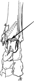
6. Twist both ends of wire together to form a figure eight. Tighten the wire just enough to compress the fracture (see Fig. 111-1).
7. Cut off excess orthopedic and Kirschner wires. Bend the Kirschner wires over and embed them in the soft tissues to trap the orthopedic wire loop and minimize soft tissue irritation.
a. In the treatment of tibial tubercle fractures, if the animal still has significant growth potential, do not use tension band wires because this procedure can induce premature physeal closure. If necessary, use additional Kirschner wires oriented perpendicular to the physis for fixation stability.


