Chapter 107 Fractures of the Pelvis
Pelvic fractures in dogs and cats are extremely common. Severe trauma, such as motor vehicular accidents, is usually the cause of pelvic fractures. Concurrent injuries to other body systems, including life-threatening injuries, are also common and must be identified and treated in a timely fashion. Several specific types of injury occur in the pelvis, including sacroiliac luxation, fractures of the non-articular portions of the pelvis, and articular fractures. Specific management protocols for each are described below.
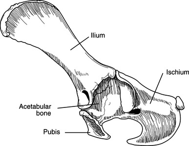
ANATOMY
DIAGNOSIS
Concurrent Thoracic and Abdominal Injuries
Pelvic Injuries
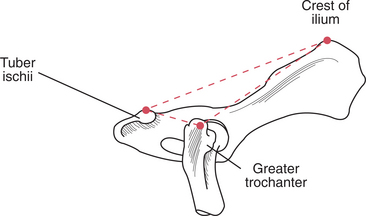
Figure 107-2 Diagram illustrating the normal position of the greater trochanter, iliac crest, and ischiatic tuberosity.
(From Fossum TW: Small Animal Surgery, 2nd ed. St Louis: Mosby, 2002.)
TREATMENT
Non-surgical Treatment
Nursing Care
Surgical Treatment
Indications
Surgical treatment is indicated for the following injuries:
FRACTURES OF THE ILIUM
Preoperative Considerations
Surgical Procedure
Equipment
Technique
Stay updated, free articles. Join our Telegram channel

Full access? Get Clinical Tree


