Chapter 5 Fishes
Case 5.1
Clinical history
• A large, black swelling was visible on the first gill arch on the right hand side
• This female had a relatively recent history of not brooding her egg clutches to full term
Physical examination
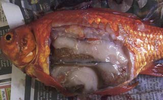
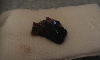
The fish was netted and the gills briefly examined by alternately lifting the operculae with the fish restrained in the net. A dark mass could be seen associated with the gills on the right hand side (Fig.5.1).
Clinical diagnosis examination
 1. List your differential diagnoses.


Therapy
 2. What treatment options are available to you?
Treatment options are limited but would include:
• Biopsy and appropriate treatment for non-neoplastic conditions
• Surgical debulking. Possible but risky in view of the potentially highly vascular tissue involved
• Surgical debulking followed by weekly cisplatin injections may be considered for skin tumours, but again this mass is in a very vascular area and surgical debulking and chemotherapy may be highly risky.
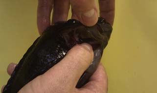
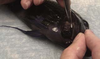
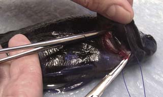
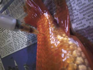
It was elected to remove the mass surgically. This was achieved by resection of the first branchial arch. This was clamped at either end and ligated with PDS (Fig.5.3). Antibiotic cover was provided with an intramuscular injection of 1 mg marbofloxacin. Recovery involved placing the cichlid back into water that did not contain MS222.
The mass was some 2.0 cm in diameter and was submitted for histological examination (Fig.5.4).
Discussion
A variety of gill neoplasms have been recorded including papillomas, squamous cell carcinomas, branchioblastomas and chondromas (Childs and Whitaker 2001). Sarcomas have been well described in a variety of fish (Earnest-Koons etal. 1996, Lewbart etal. 1998, Schmale etal. 2002), although their presence in cichlids does not appear to have been widely reported. In the walleye, dermal sarcomas are due to a retroviral infection and can be transmitted (Earnest-Koons etal. 1996) – interestingly, other sarcomas in fish appear to be linked to viral agents or at least a virus-like agent (Schmale etal. 2002),
Childs S., Whitaker B.R. Respiratory disease. In: Wildgoose W.H., ed. BSAVA Manual of Ornamental Fish. second ed. Gloucester: British Small Animal Veterinary Association; 2001:145.
Earnest-Koons K., Wooster G.A., Bowser P.R. Invasive walleye dermal sarcoma in laboratory-maintained walleyes Stizostedion vitreum. Dis. Aquat. Organ.. 1996;24:227–232.
Lewbart G.A., Spodnick G., Barlow N. Surgical removal of an undifferentiated abdominal sarcoma from a koi carp (Cyprinus carpio. Vet. Rec.. 1998;143(20):556–558.
Schmale M.C., Gibbs P.D.L., Campbell C.E. A virus-like agent associated with neurofibromatosis in damselfish. Dis. Aquat. Organ.. 2002;49:107–115.
Case 5.2
Post-mortem diagnosis examination
Initially a skin scrape was taken, and then both fish were euthanased with intravenous pentobarbitone (Fig.5.5). A post-mortem examination was then undertaken. The first goldfish weighed 179 g and had a standard length of 152 mm. The results for this first goldfish are:

Gill squash
 Huge numbers of these parasites were found
 Normal gill lamellar architecture disrupted. Significant clubbing of lamellae
 1. What are these parasites likely to be?
 2. Why is it important to distinguish between Dactylogyrus and Gyrodactylus?
On examination of the coelomic cavity, the fish was revealed to be female with well-developed ovaries showing evidence of asynchronous egg development. The carcass was very fatty and the caudal kidney appeared very soft with a loss of normal texture. The spleen appeared enlarged (Fig.5.6).