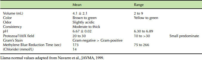
Once collected, the fluid should be placed in a clean vial and either analyzed immediately or kept at body temperature until timely analysis can be performed (Table 40.1).
Table 40.1 Normal first compartment fluid values in llamas.
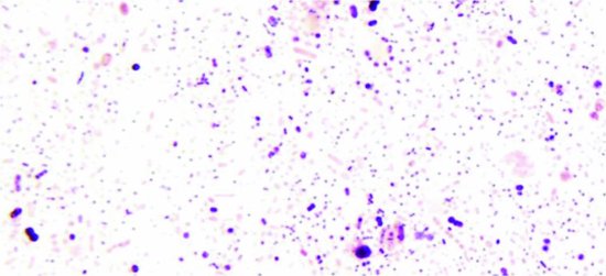
C-1 fluid pH is best measured using a pH meter, because they are less affected by the color of the fluid than litmus papers. A drop of fluid may be placed on a warm glass slide and examined using a microscope. On low power, numerous protozoa should be seen moving rapidly across the slide. (See Figure 40.2; Video 40.1: Protozoa low power.) Three sizes of protozoa are normally present (small, medium, and large) and can be easily discerned using high magnification (Video 40.2: Protozoa high power). The large protozoa are the most sensitive to lactic acidosis and will be the first to be lost in cases of first compartment acidosis. This slide may then be dried and Gram stained to assess ratios of Gram-positive and Gram-negative bacteria. (See Figure 40.3.) Recall that Gram’s staining was designed for use on pure cultures of bacteria, and staining can be misleading with a very heterogenous population such as rumen flora.

Figure 40.2 Iodine-stained protozoa. These can be observed in fresh fluid rapidly moving, and a population of small, medium, and large should be seen.
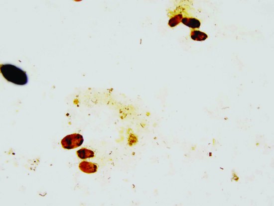
Figure 40.3 Gram stained first-compartment fluid. Many “artifacts” of C-1 fluid take up stain and make Gram staining difficult to interpret.
Stay updated, free articles. Join our Telegram channel

Full access? Get Clinical Tree


