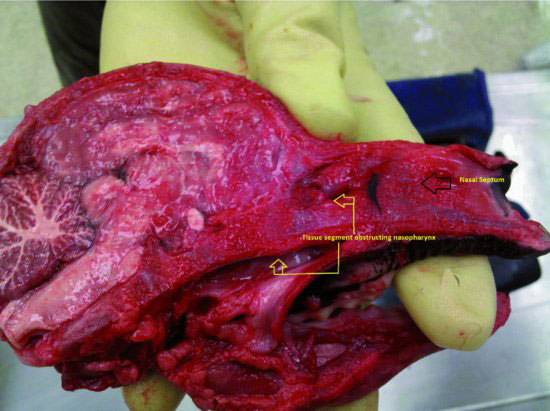
Figure 34.1 Sagittal section through the head of an alpaca cria demonstrating the atretic tissue occluding the nasopharynx.

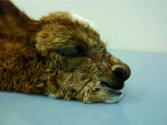
Figure 34.2 Obstruction of the nasopharynx results in breathing patterns associated with flaring of the nostrils and opening of the mouth.

EQUIPMENT NEEDED
The following equipment is needed: 12-mL syringe, red rubber feeding tube (10 to 14 French), loose cotton, mirror, normal saline, pediatric bronchoscope (optional), radiographic equipment (optional), and radiopaque contrast material (optional).
RESTRAINT/POSITION
Sternal recumbency (cushed posture) or lateral recumbency is used. Sedation is not recommended unless orotracheal intubation is also done.
TECHNICAL DESCRIPTION OF PROCEDURE/METHOD
Crias suspected of being affected with choanal atresia can be difficult to make an accurate diagnosis because of the small size of the head and nasopharynx and because of the location of the obstruction. The obstruction is normally located 8- to 10-cm caudal to the nares and at the level of the medial canthus of the eye. If clinical signs are consistent with choanal atresia, then confirmatory tests should be done before euthanasia is opted. There are six readily performed clinical tests to determine if complete obstruction of the nasal passageway is present. These tests are most useful when complete (bilateral) choanal atresia exists. In the rare cases of unilateral choanal atresia, false test results can easily occur with the exception of endoscopy.
Follow these steps:
Stay updated, free articles. Join our Telegram channel

Full access? Get Clinical Tree


