However, moderate to severe disruption of the tympanic bullae, as seen on CT scans, has been diagnosed with little to no outward clinical signs in the patient. Otitis media/interna in llamas and alpacas is thought to develop after ascending infection from the external auditory canal. However, these infections may also arise from the nasopharynx by ascension through the Eustachian tubes. Herd outbreaks of otitis externa have been observed when alpacas were randomly misted with water to keep them cool during the heat of summer. Herd outbreaks of otitis interna/media have been diagnosed after infestation with spinose ear ticks.
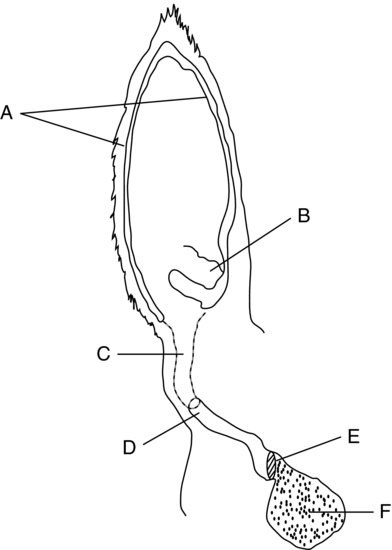
Difficulty arises when examining the ear and ear canal in camelids. Llamas and alpacas resist handling of the ear especially if there is a history of frequent use of the ear during manual restraint. This procedure may require sedation. Otoscopic examination is very limited due to the anatomical shape and size of the ear canal (Fowler and Gillespie, 1985) (Figure 27.2). Flexible endoscopes, radiographs, and computed tomography are often helpful to evaluate problems involving the ear canal, internal ear, and tympanic bullae.
Figure 27.2 Sketch diagram (cutaway anterior view) of the structures of the external, middle, and inner right ear: (A) the cartilage of the external pinna, (B) the area of the conchal eminence, (C) the lateral vertical canal, (D) the beginning of the bony horizontal canal, (E) the tympanic membrane, and (F) the tympanic bulla.

EQUIPMENT NEEDED
An otoscope with a long slender cone will be needed. Optional equipment may include a flexible pediatric endoscope (<6 mm diameter), radiographic equipment, and computed tomography. In llamas or alpacas suspected of having congenital ear defects, otic biopsy may be performed for histopathologic examination. A 2-mm to 4-mm diameter skin biopsy punch or a swine ear-notching tool is an efficient method for specimen collection.
RESTRAINT/POSITION
Camelids resist examination and handling of the ear for examination, especially when the ears are painful or inflamed. Sedation is usually necessary. Butorphanol (0.1 mg/kg IV) and/or xylazine (2 to 4 mg/kg IV) are sufficient to facilitate most otic examinations.
TECHNICAL DESCRIPTION OF PROCEDURE/METHOD
First, view the external pinna of the ear for clinical signs of trauma (laceration, aural hematoma), alopecia, infection, or parasite infestation. The integrity of the cartilage should be evaluated in neonates in conjunction with physical exam and signalment parameters to attempt to differentiate weak cartilage from congenital defects (Fowler, 1998) (Figure 27.3).
Figure 27.3 Photograph of a newborn cria that was showing signs of prematurity. Note the lax cartilage of the external pinna, which resolved within a few days of birth. Congenital cartilage laxity is more pronounced and does not resolve.

Stay updated, free articles. Join our Telegram channel

Full access? Get Clinical Tree


