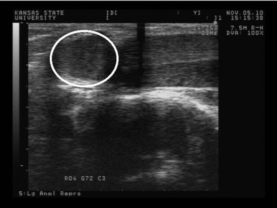
Figure 61.1 The bulbourethral glands are located immediately dorsal to the ischial arch and cranial to the anal sphincter. The glands are recognized by their spherical shape and homogeneous echotexture.

Prostate Glands
The bilobed prostate glands are located immediately caudal to the attachment of the vas deferens to the pelvic urethra caudal to the trigone region of the bladder (Figure 61.2). These glands are oval in shape and extend dorsal and lateral from the dorsal surface of the pelvic urethra. These glands have a broad base and can be difficult to measure accurately, but the prostate glands should be symmetrical and ultrasound measurements of their dimensions within 10% of the contralateral gland (Table 61.1). The prostate can, occasionally, be evaluated by palpation. More often, ultrasound examination via the rectum is required. High frequency ultrasound probes (7.5 to 10 MHz) are desirable for this evaluation so that higher resolution images can be obtained. In alpacas, a rectal linear ultrasound probe can usually be inserted through the anal sphincter far enough to visualize the glands. In llamas, the probe may need to be controlled via rectal palpation or fitted to a rigid extender (50-cm long), such as a molded PVC tube, so that the probe can be inserted through the rectum far enough for complete examination. These glands should be assessed for symmetry, size, and tissue echotexture. The glands should be homogeneous in nature, and the prostatic duct can often be visualized coursing through the center of the gland and connecting to the pelvic urethra (Figure 61.3). The pelvic urethra and bladder also are examined at this time. Camelids, especially alpacas, can suffer prostatitis, prostatic abscesses, prostatic cysts, and idiopathic prostatomegaly.
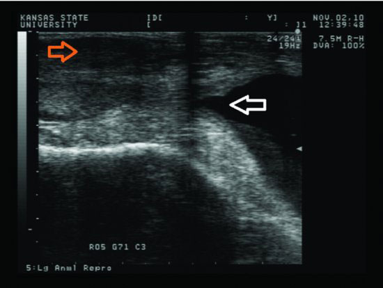
Figure 61.2 The prostate glands are located within the pelvic canal and caudal to the trigone of the bladder and attachment of the vas deferens. The trigone (open white arrow) is most easily recognized when the bladder contains a sufficient volume urine. The prostate is homogeneous in echotexture and caudal to the trigone.

Stay updated, free articles. Join our Telegram channel

Full access? Get Clinical Tree


