Chapter 22 Erythrocytes, Leukocytes, and Platelets
ERYTHROCYTE DISORDERS
Anemia: Overview
Clinical Signs
Principles of Diagnosis
Several tests can be used to document and characterize the anemia morphologically or etiologically.
History and Physical Examination
Use the history and physical examination to determine the following:
Complete Blood Count (Table 22-1)
Table 22-1 HEMATOLOGY REFERENCE RANGES FOR DOGS AND CATS*
| Parameter | Canine | Feline |
|---|---|---|
| PCV or Hct (%) | 37–55 | 30–45 |
| Hemoglobin (g/dl) | 12–18 | 8–15 |
| RBCs (×106/μl) | 5.5–8.5 | 5–10 |
| MCV (fl) | 60–75 | 40–55 |
| MCHC (g/dl) | 32–36 | 30–36 |
| Reticulocytes (×103/μl) | <80 | <60 aggregate |
| Platelets (×103/μl) | 200–900 | 300–700 |
| WBCs (×103/μl) | 6–17 | 6–18 |
| Segmented neutrophils (×103/μl) | 3–12 | 3–12 |
| Band neutrophils (/μl) | 0–300 | 0–300 |
| Lymphocytes (/μl) | 1000–5000 | 1500–7000 |
| Monocytes (/μ) | 150–1350 | 50–850 |
| Eosinophils (/μl) | 100–1250 | 100–1500 |
| Basophils (/μ) | <100 | <100 |
| Plasma protein (g/dl) | 6.0–8.0 | 6.0–8.0 |
| Fibrinogen (mg/dl) | 200–400 | 150–300 |
Hct, hematocrit; MCHC, mean corpuscular hemoglobin concentration; MCV, mean corpuscular volume; PCV, packed cell volume; RBCs, red blood cells; WBCs, white blood cells.
Source: Purdue University, Veterinary Clinical Pathology Laboratory.
* These values are only meant as a guide; individual laboratories may vary in their ranges, depending on instrumentation and regional differences in animal populations.
Reticulocyte Count

Fecal Examination and Urinalysis
These tests are performed to determine sources of blood loss and function of the kidney.
Bone Marrow Evaluation
Bone Marrow Aspiration Biopsy Technique
Principles of Transfusion Therapy
Therapy depends on the etiology of the anemia. Rapid decreases in PCV warrant replacement of whole blood. However, slow daily decreases in PCV of 1% to 3% may not cause clinical signs of dyspnea or weakness. Whole blood transfusion is discussed here, and blood component therapy is discussed in Chapter 23.
Cross-matching
Whole Blood Transfusion
Hemolytic Anemia
Causes of hemolytic anemia include congenital abnormalities, immune-mediated destruction, infections, chemical or toxic agents, mechanical fragmentation, and hypophosphatemia. The net effect of hemolytic loss of erythrocytes is often a very strong to moderate regenerative response (Fig. 22-1). However, in some cases, the anemia may occur so rapidly that the animal may have too little time to mount a regenerative response by the time the condition is recognized.
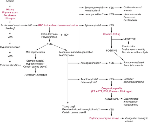
Figure 22-1 Diagnostic approach to common regenerative anemias in dogs and cats.* See Figures 22-2 and 22-3. (APTT, activated partial thromboplastin time; FDP, fibrin degradation product; PT, prothrombin time.)
Congenital Erythrocyte Abnormalities
Pyruvate Kinase Deficiency
Clinical Signs
Phosphofructokinase Deficiency
Diagnosis.
Hereditary Stomatocytosis
Feline Porphyria
Infectious Causes of Hemolysis
Hemoplasmosis
Diagnosis
Babesiosis
Diagnosis
Initial diagnosis is made by examination of a blood smear.
Chemical or Toxic Injury of Erythrocytes
Oxidant-Induced (Heinz Body) Anemia
Snake Venom Toxicity
Mechanical Fragmentation of Erythrocytes
Heartworm Disease
Disseminated Intravascular Coagulopathy
Nonregenerative Anemia
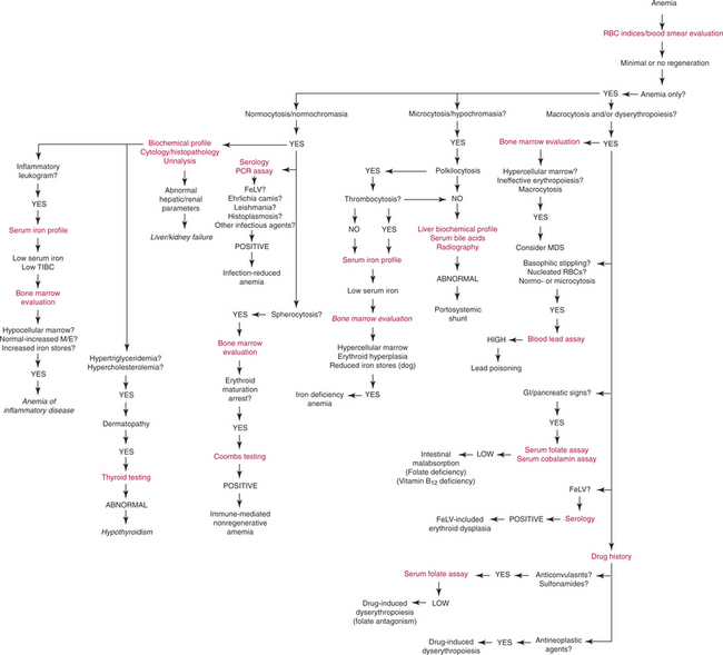
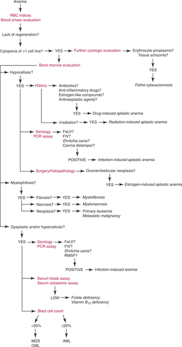
Nonregenerative anemia generally is related to direct toxicity of erythroid precursors in the bone marrow or secondary suppression of erythropoiesis (Fig. 22-2). Often neutrophils and platelets also are affected (Fig. 22-3).
Infectious Agents
Feline Leukemia Virus
Diagnosis.
Diagnosis is based on positive enzyme-linked immunosorbent assay (ELISA) or IFA test results (see Chapter 8). Bone marrow aspirate smears or core biopsy sections indicate decreased cellularity with increased fat infiltration when the infection is not associated with a proliferative leukemia.
Treatment.
Treatment varies depending on concurrent conditions such as neoplasia, hemoplasmosis, FIV, or feline infectious peritonitis (FIP). Supportive care, in the form of blood transfusions (see “Principles of Transfusion Therapy”), antibiotics, appetite stimulants, and anabolic steroids, is often necessary (see Chapter 8 for additional information on FeLV).
Ehrlichiosis and Anaplasmosis
Ehrlichia spp. and Anaplasma spp. are tick-transmitted rickettsial infections that produce chronic polysystemic disease in dogs and cats associated with hyperglobulinemia, thrombocytopenia, mild to moderate non-regenerative anemia, and sometimes leukopenia or pancytopenia in the later stages. The epidemiology, clinical manifestations, diagnosis, and treatment of these infections are described in Chapter 17.
Stay updated, free articles. Join our Telegram channel

Full access? Get Clinical Tree


