CHAPTER 52 Epizootic Bovine Abortion (Foothill Abortion)
HISTORY AND ETIOLOGY
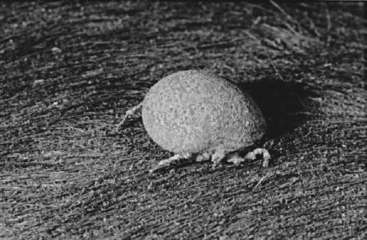
Epizootic bovine abortion (EBA), also known as foothill abortion, is a distinct, tick-transmitted disease of pregnant cattle that graze foothill and mountain pastures of California and adjacent states.1 Endemic areas often are bushy or wooded regions, typically dry chaparral or oak-studded environments from 500 to 7000 feet above sea level, and are the habitat of the argasid tick vector Ornithodoros coriaceus2 (Fig. 52-1). Heifers confined to permanent (irrigated) pastures within endemic areas rarely if ever experience EBA.

Fig. 52-1 Adult engorged Ornithodoros coriaceus tick, the vector of epizootic bovine abortion.
(Courtesy of M. Oliver.)
A causative agent for EBA has been sought for more than 50 years.3 Research through the mid 1980s suggested several different agents, including chlamydiae (Chlamydophila), viruses, and spirochetes; however, recent breakthroughs, using molecular techniques, chemical and immunohistologic stains, and other methods, have implicated a bacterial agent.3–6 Although the disease has a limited geographic distribution, it is a major cause of abortion, neonatal mortality, and economic losses in endemic areas. It also is of interest to the veterinary scientist because of its unusual presentation, long latent period, and pathologic features.
NATURAL HISTORY
In the 1960s, the geographic distribution of the soft-bodied tick, O. coriaceus (see Fig. 52-1), was shown to coincide with the known areas in which EBA had been reported.2 The most common hosts of this ectoparasite are deer and cattle. Experimental feeding of the tick on susceptible pregnant heifers was shown to reproduce the disease.2,7 On the basis of this and other work, the role of O. coriaceus as a vector of the EBA agents has been accepted. Various agents have been isolated from this tick, but none of these agents has reliably reproduced EBA.3 Experimental work has shown that the thymus of EBA-aborted fetuses reliably transmits the disease when extracts are inoculated into susceptible pregnant heifers. In recent studies, the “thymic agent” was shown to be nonfilterable (e.g., infectivity was removed by filtration); it also was destroyed by sonication, and its pathogenicity was ablated by antibiotic treatment.4 Although no agent has been reliably cultivated from the thymus, molecular techniques that amplify microbial nucleic acids (polymerase chain reaction [PCR] assays) identified a deltaproteobacterial agent, specifically of the Myxococcales order.5 This order is notorious for failure of organisms to grow in in vitro culture. So, although the demands of classic Koch’s postulates have not been met, a majority of guidelines that have been suggested for DNA sequence–based determination and incrimination of a presumptive microorganism in disease have been satisfied.8
CLINICAL SIGNS AND FINDINGS
Heifers or cows that have been either naturally or experimentally exposed to EBA show no overt signs of illness at any time after exposure. Minor elevations in total leukocyte counts about 1 week following tick feedings have been noted. The interval from exposure to abortion is unusually long, typically 3 to 4 months, with nearly all abortions coming in the third trimester. When fetuses are expelled, they usually are in a relatively fresh (i.e., nonautolytic) state. Serum from aborted EBA fetuses shows markedly elevated immunoglobulin concentrations, especially immunoglobulin G (IgG) (150–800 mg/dl; geometric mean, approximately 300 mg/dl), and in some instances, the immunoglobulin levels can approach those of colostrum-fed neonates (>1 g/dl).7,9 We are not aware of another disease entity that routinely stimulates such high levels of bovine fetal immunoglobulin.
Stay updated, free articles. Join our Telegram channel

Full access? Get Clinical Tree


