RESTRAINT/POSITION
The procedure can be completed with the patient standing or in sternal recumbency. Sternal positioning has the benefit of widening the epidural space for catheter placement. Sedation may be necessary in some cases.
TECHNICAL DESCRIPTION OF PROCEDURE/METHOD
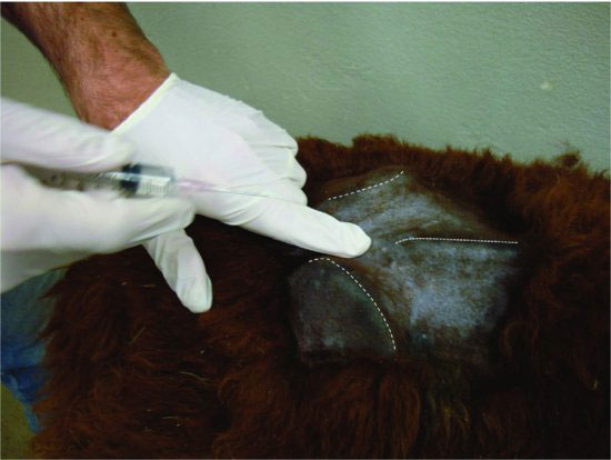
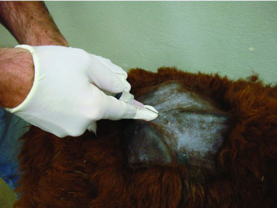
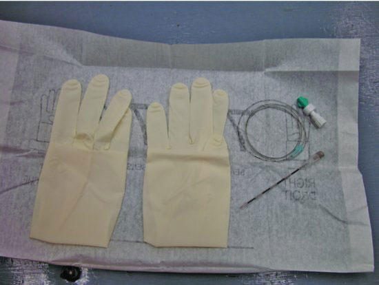
The area of catheterization must be clean and aseptic techniques used. Clip and surgically prep the lumbar sacral area for catheter placement. (See Figure 21.2.) Inject 1 to 2 mL of 2% lidocaine in the subcutaneous tissues, but avoid puncture of the ligamentum flavum and dura, then aseptically prepare the area again. (See Figure 21.3.) Place the materials needed for catheter placement in a sterile field. (See Figure 21.4.)
Figure 21.2 The clinician’s index finger is placed on the location of the lumbar-sacral space approach. The tuber coxae of the pelvis and the dorsal spinous processes of the sacral vertebrae are marked with dashed white lines.

Figure 21.3 One to two milliliters of 2% lidocaine are injected into the subcutaneous tissues en route to the epidural space.

Figure 21.4 The materials needed for epidural catheterization are placed in a sterile field for efficient use. It is very important to be sterile in placement of the catheter.

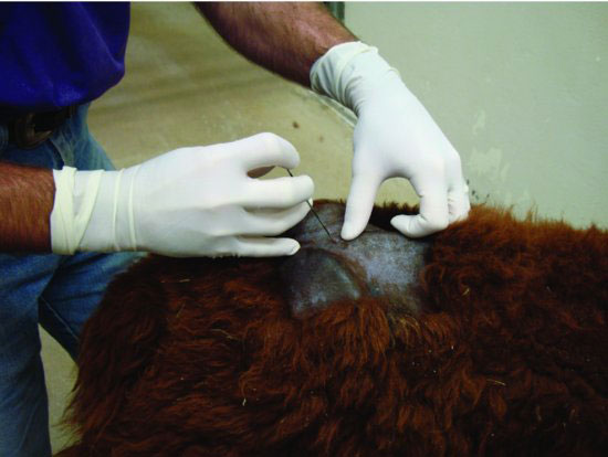
Insert the Tuohy needle through the skin to the epidural space. (See Figure 21.5.) Advance the needle toward the epidural space slowly so that you do not puncture the dura into the subarachnoid space. When you feel the epidural space has been reached, remove the stylette and test for proper placement. If spinal fluid is observed in the hub of the needle, the needle is positioned too deep and should be retracted. Methods to test for epidural placement include the following: (1) saline solution freely flows from the hub of the needle into the space, or (2) the epidural catheter is easily advanced. (See Figure 21.6.) The epidural catheter placement will be met with some resistance even when correctly entering the epidural space. Resistance to advancement must be interpreted carefully so as to minimize repositioning of a correctly placed needle.
Figure 21.5 The Tuohy needle is placed and advanced toward the epidural space. Be sure the curve of the needle faces forward. Do not insert the needle too deep (refer to text for discussion).

Stay updated, free articles. Join our Telegram channel

Full access? Get Clinical Tree


