Fig. 1.
Electrode placement on hippocampal slice. Recording electrode on the cell layer of CA1 and stimulating electrode in the stratum radiatum of CA3.
Record basic neurotransmission by stimulating the Schaffer collateral pathway using a pulse range from 10 to 300 μA at a rate of 0.05 Hz to find the maximum response.
To examine LTP, record the baseline for 30 min by stimulating the pathway with the pulse intensity that gives 30–40% of the maximum EPSP at the rate of 0.05 Hz.
Induce LTP by stimulating the CA3-CA1 pathway with high frequency stimulation (HFS) consisting of 4 trains of 100 pulses at 100 Hz with an inter-train interval (ITI) of 1 min.
Record the post-HFS response for 2 h.
The signal is amplified, digitized and then acquired using a personal computer with Clampex 8 software running.
2.4 Data Analysis
Use clampfit 9.2 to quantify the slope of the rising phase of the EPSP and the amplitude of population spike.
Normalize all the data to baseline which is set as zero.
Compare the mean of post-HFS response at different time points among the groups to look for significant differences in the degree of potentiation between the groups.
Assess the mean response induced immediately after HFS to compare the induction phase of LTP between groups.
Statistical tests, such as one-way analysis of variance or Student’s t-test can be used.
3 Variations in Electrophysiological Recording
3.1 Variation in HFS
3.2 When to Perform Electrophysiology After SAH
LTP after experimental SAH can be examined at different time points after experimental SAH.
3.3 Alternative Methods to Examine Hippocampal Function
Using field potential recording, short-term plasticity of hippocampal synapses could be examined. The most typical short-term plasticity method is pair-pulse facilitation at the CA3-CA1 pathway in which a pair of identical pulse stimulation with inter-pulse interval of 50 ms is applied by the stimulating electrode. The potentiation of the second response over the first one would mean enhanced short-term plasticity.
Whole cell patch clamp technique is another alternative method to obtain specific information about hippocampal cell function by studying the ion channels and associated receptors, as well as the other components of the cellular machinery that control cell function. This method is more technically demanding. Field potential recordings are simpler, easier to learn and set up, the equipment is less costly but they provide a relatively general overall assessment of populations of cells in the recorded regions.
4 Assessment of Hippocampal Function by Behavior Testing Using Morris Water Maze
Many procedures have been developed that use escape from water to stimulate learning and memory in animals (19, 20). Water maze tasks have been quite useful in evaluating the effects of experimental lesions and drug effects in rodents by measuring spatial learning. The Morris water maze (MWM) was developed in 1981 and further characterized in 1984 (21). It is widely accepted and is extensively used to examine a variety of aspects of hippocampal function, such as spatial memory, as well as many other aspects of neurocognitive function (22).
The advantages of the MWM are that it allows for examining a variety of performance and learning assessments without extensive pretraining since animals are natural swimmers and learn the water-escape tasks easily (21). In addition, previously used aversive procedures, such as food deprivation, difference in water temperature, and electric shock techniques to motivate learning are not required. Furthermore, the water provides a control for olfactory cues, video tracking devices can specifically identify the motor or learning deficits by eliminating the non-mnemonic behaviors, such as thigmotaxis, and relearning experiments can be performed by changing the platform location (23). Disadvantages of the MWM are the relatively high cost of apparatus, requirement for a large-enough space and a quiet environment to conduct experiments. In addition, immersion into water may cause endocrinological or other stress effects which may interact with experimental manipulations and results in uncontrolled ways.
4.1 Materials and Instruments
Large circular pool made of plastic and painted black (e.g., diameter: 180 cm, height: 76 cm for rats and diameter: 120 cm, height: 76 cm for mice).
Water to fill the pool.
Powdered milk or nontoxic paint to opaque the water (if needed).
Highly reflective paint
A clear plexiglass platform (10 × 10 cm)
Unique geometric images made of cardboard.
Flag made of plastic tube and tape.
Black curtains
Video camera
Stopwatch
Automatic tracking system (Poly Track System, San Diego Instruments, San Diego, CA).
4.2 Preparation of Water Maze
All behavioral tests should be performed at the same time of the day.
Circular pool is filled with water to a depth of 40 cm at room temperature (26 +/- 1°C).
Four equally spaced points around the circumference of the pool are arbitrarily designated North (N), South (S), East (E), and West (W), and thus four quadrants are established (NW, SW, NE, and SE), and eight locations all together.
The area between an arbitrary inner circle (20 cm from the wall) and the pool wall is designated as the wall area @ defining thigmotaxis.
Three to six unique visual cues, such as highly reflective geometric images, are placed on the walls.
Black curtains are used to hide extra-maze cues, such as the experimenter and other animal subjects for some tests.
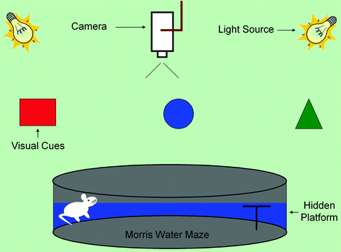
A video camera is mounted in the center of the ceiling above the pool to monitor the animal’s behavior through an automatic tracking system (Poly Track System, San Diego Instruments, San Diego, CA, Fig. 2). Tracking is achieved due to contrast between a white animal and black background or vice versa with suitable reflected lighting.

Fig. 2.
Morris water maze setup illustrating locations of pool, camera, light source, visual cues, and platform.
4.3 Spatial Acquisition Task (Hidden Platform Test)
This task is performed over 6 consecutive days to examine spatial memory.
Each animal is subjected to sixteen 60 s trials of hidden platform with inter-trial interval of 10 s each day. The first trial on day 1 is excluded from data analysis to allow the animal to acclimate to the experimental conditions.
Place the platform approximately 2.0 cm below the surface of water in the center of one of the quadrants to make it invisible for animals. The location of the platform is changed to a novel location every second day.
Place the animal in water facing the pool wall with the starting point randomized among the rest of the seven locations to exclude the shortest path to the platform.
If the animal is unsuccessful in finding the platform within 60 s, guide it to the platform. Allow the animals to stay on the platform for additional 10 s after they climb on or are guided to the platform.
< div class='tao-gold-member'>
Only gold members can continue reading. Log In or Register to continue
Stay updated, free articles. Join our Telegram channel

Full access? Get Clinical Tree


