Electrocardiography
Basic Information 
Overview and Goal(s)
• An electrocardiogram records the depolarization and repolarization process in atria and ventricles during the cardiac cycle.
• Electrocardiography is used to evaluate cardiac rate and rhythm. It allows the diagnosis of the nature of a dysrhythmia (eg, origin of ectopy, type of conduction disturbance).
Indications
Equipment, Anesthesia
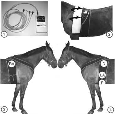
• Electrocardiograph (ECG machine) to make the recording. This can be a large or small (handheld) recorder, or a recorder that transmits data wirelessly to a computer with appropriate software (Figures 1 and 2).
• Cable to connect the machine to the electrodes on the patient.
• Electrodes: Best results are obtained with self-adhesive (equine) electrodes. These are well tolerated, easy to use, and produce few artifacts. Crocodile clamps are more painful and produce more movement artifacts, although they are also often used.
• The ECG trace can be displayed on a screen, recorded on paper, or digitally recorded (digital media, computer).
• A single lead recording is satisfactory in most cases. However, multiple lead recordings may be helpful in some situations.

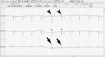
FIGURE 2 Example of an ECG obtained from the system described in Figure 1. Electrodes are placed as indicated in figure 1 to record three leads in this horse. Leads I and II produce the largest deflections. However, ventricular premature complexes are hard to differentiate from the normal complexes on lead II (*). The difference is more obvious on lead I (arrowheads) but most easily seen on lead III (arrows), which shows the smallest deflections.
Preparation: Important Checkpoints
• Check recording unit for paper supply, batteries (ink: rarely used).
• Take measures to protect equipment with animals at risk for collapse.
• Take measures to protect equipment when horses are free in a box or paddock during long-term recording (eg, to prevent damage during recumbency or rolling).
Stay updated, free articles. Join our Telegram channel

Full access? Get Clinical Tree


