Chapter 7 Domestic Rabbit (Oryctolagus cuniculus)
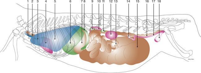
Figure 7-1, A Anatomic drawing (view of the left side) of viscera of the thorax and abdomen of an adult female rabbit.
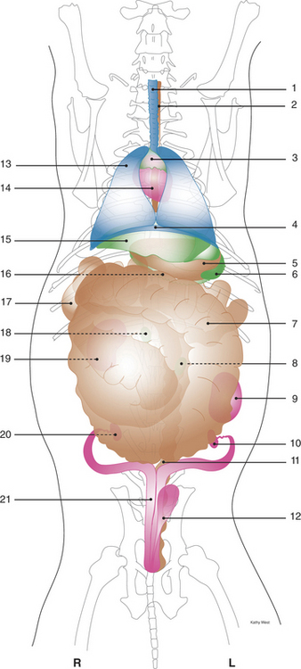
Figure 7-1, B Anatomic drawing (ventrodorsal view) of viscera of the thorax and abdomen of an adult female rabbit.
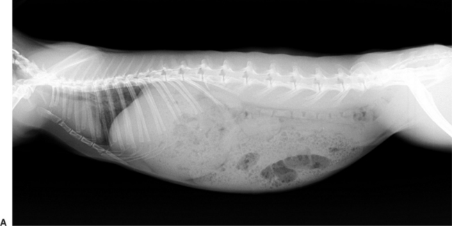
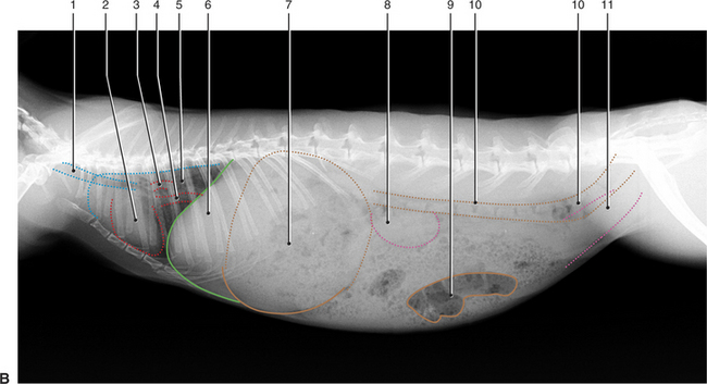
Figure 7-2, A and B Type of animal: Rabbit
Type of study: Viscera of thorax and abdomen
Projection: Laterolateral (right lateral recumbency)
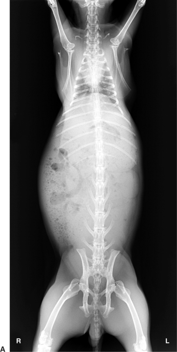
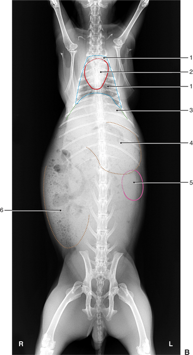
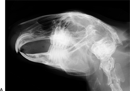
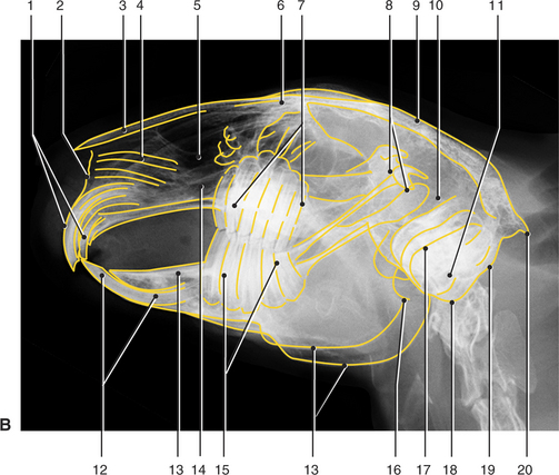
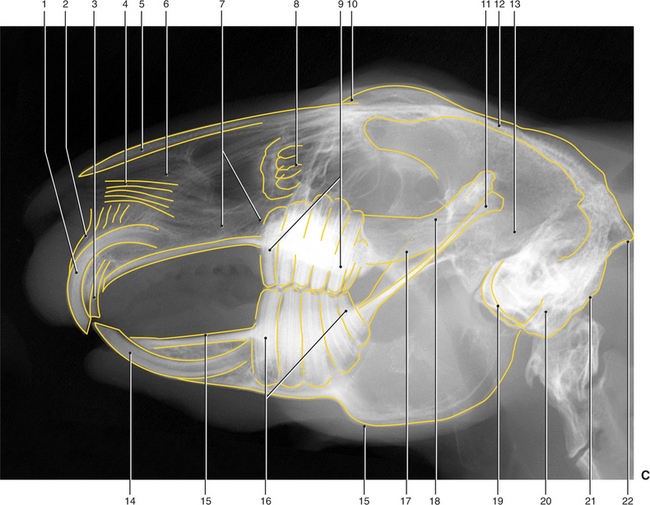
Figure 7-4, C Type of animal: Rabbit
Type of study: Magnification study of head
Projection: Laterolateral (right lateral recumbency)
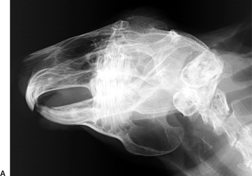
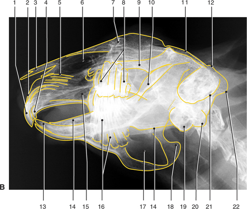

Figure 7-5, C Type of animal: Rabbit
Type of study: Magnification study of head
Projection: Oblique (10 degree) ventrodorsal
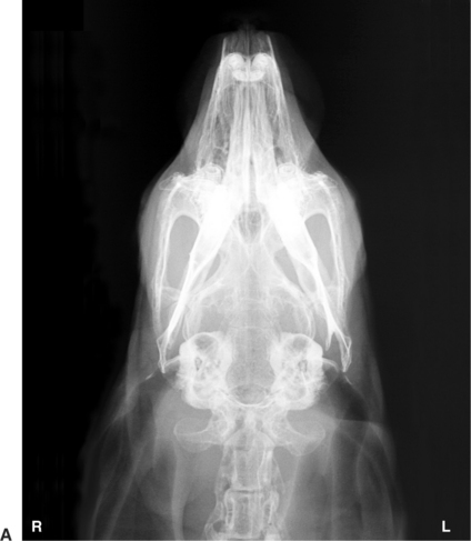
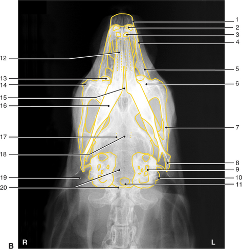
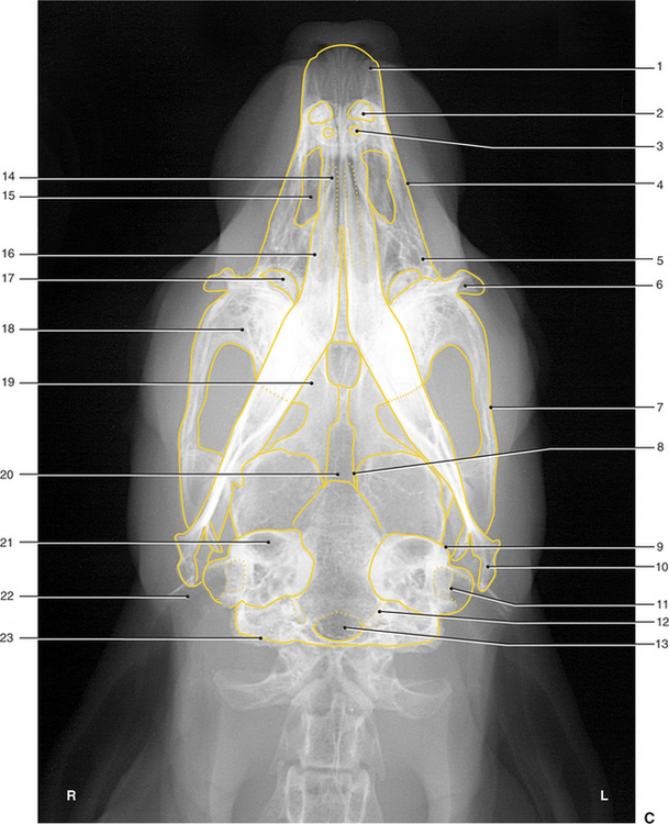
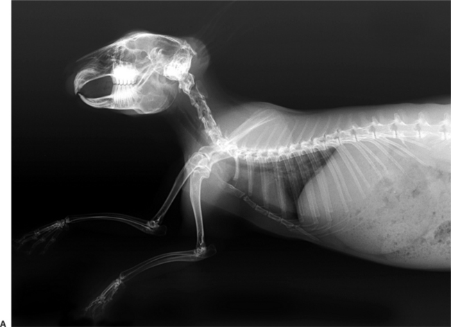
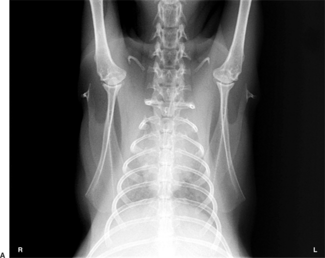
Figure 7-7, A Type of animal: Rabbit
Type of study: Cervical and thoracic vertebral column
Projection: Laterolateral (right lateral recumbency)
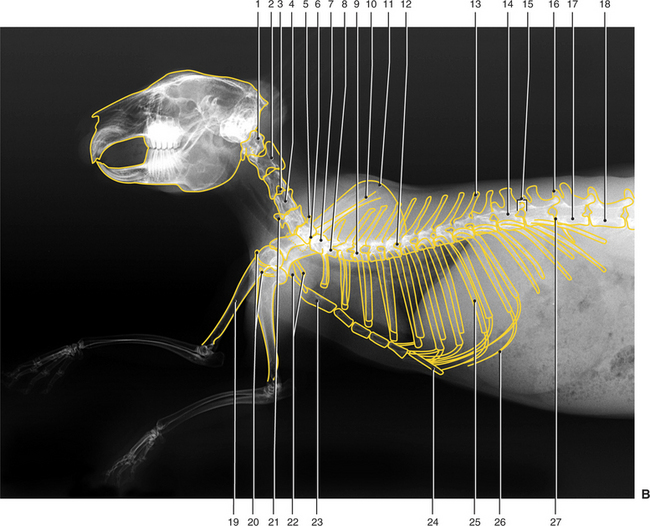
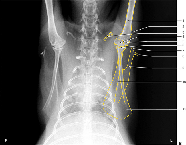
Figure 7-7, B Type of animal: Rabbit
Type of study: Cervical and thoracic vertebral column
Projection: Laterolateral (right lateral recumbency)
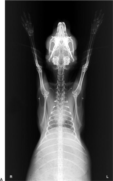
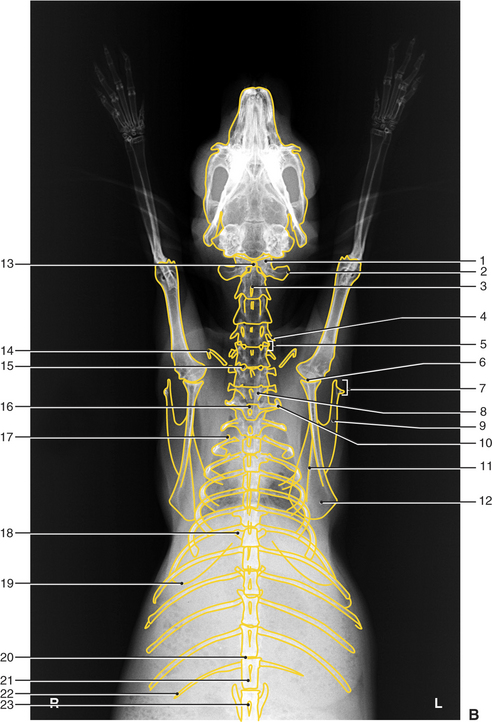
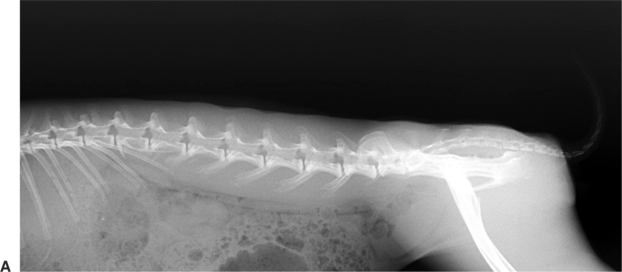
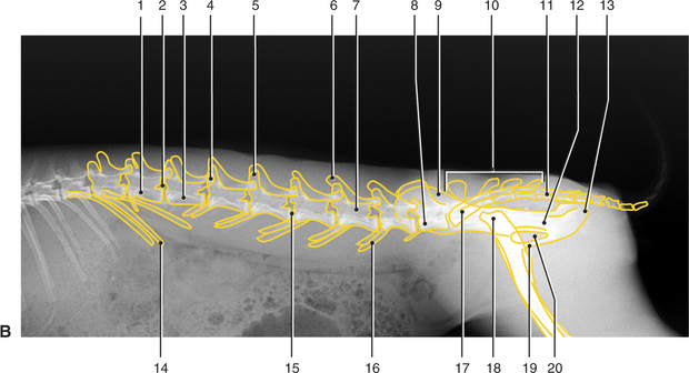
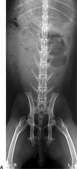
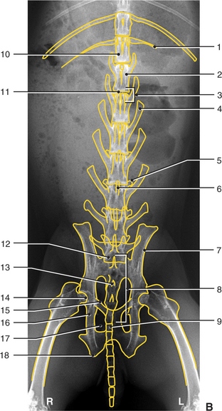
Figure 7-9, A and B Type of animal: Rabbit
Type of study: Lumbar, sacral, and caudal vertebral column
Projection: Laterolateral (right lateral recumbency)