Chapter 63 Disorders of the Claw
INTRODUCTION
Onychology (the study of nails) is an area of veterinary dermatology that has only recently become the focus of more detailed study. In the past decade, several studies have shed light on the etiology, diagnosis, and treatment of claw disease in the dog. Some relevant terms are explained in Table 63-1. A thorough diagnostic approach is essential, as treatment is successful only when the cause of the disease is known. In this chapter, a review of claw anatomy is followed by the diagnostic approach to claw disease, and finally a discussion of the treatment options for the most common disorders affecting the claw is presented.
Table 63-1 Definition of Terms Used to Describe Claw Disease
| Paronychia | Inflammation involving the folds of tissue surrounding the claw |
|---|---|
| Onychodystrophy | Malformation of the claw |
| Onychomadesis | Separation of the claw from its bed |
| Onychomalacia | Softening of the claw |
| Onychomycosis | Fungal disease of the claw |
| Onychorrhexis | Brittleness, spontaneous splitting, or breaking of the claws |
| Onychoschizia | Separation of the claw from its bed |
| Onychia | Inflammation of the claw or claw bed, resulting in loss of the claw |
| Onychogryposis | Abnormal hypertrophy and curving of the claws |
| Onycholysis | Complete loss of the claws |
ANATOMY
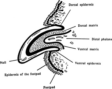
• The anatomy of the claw is shown in Figure 63-1. The superficial layers of the epidermis are modified to form the claw horn. The claw is curved and consists of a sole (the most distal portion), two laterally compressed walls (forming the axial and abaxial surfaces of the claw), and a central dorsal ridge. This ridge is thicker than the walls and sole, which maintains the pointed appearance of the claw. The coronary border (vallum) of the claw fits into the space under the ungual crest, a crescent-shaped dorsal process of the distal third phalanx. Most of the claw is formed from the coronary band, which is surrounded and hidden by the claw fold.
• The claw fold is a fold of skin at the proximal border of the claw that is continuous with the horn of the claw. Dorsally, it is a modification of haired skin. The periosteum of the distal phalanx is continuous with the dermis of the claw; these two tissues occupy the space between the bone of the distal phalanx and the claw itself.
• The corium or dermis underneath the claw epidermis and horn is often referred to as the quick. This tissue is highly vascularized, which is readily demonstrated by hemorrhage after excessive trimming of the claws.
• Any inflammation of the dermis, which is present between these two rigid structures, leads to swelling and explains why pronounced pain occurs rapidly with any disease of the canine claw matrix.
• The keratogenous zone (the basal cell layer and lower spinous cell layers) at the very proximal end of the claw is the proliferative claw matrix thought to be actively involved in producing the claw plate. Damage to the claw matrix typically results in malformation of the plate.
• The normal canine claw epithelium lacks a granular layer. The epithelium produces keratinocytes that flatten, cornify, and fuse to form the claw horn. The claw wall is curved, laterally converging and enclosing the sole distally.
ETIOLOGY
• Trauma is the most common cause of claw disease, particularly if only a single claw or a single paw is affected. Animals typically present with lameness and pain, particularly if the claw plate is loose.
• Neoplasia is a common cause of claw disease affecting single digits. A variety of tumors such as melanoma, mast cell tumors, keratoacanthoma, inverted papilloma, lymphosarcoma, fibrosarcoma, osteosarcoma, and others have been reported to affect the canine claw and claw bed. However, squamous cell carcinoma is the most common neoplasm of the distal digit and seems to be particularly frequent in large-breed dogs with black hair coats. Squamous cell carcinoma often invades the bone of the distal phalanx.
• Systemic disease is likely if multiple digits on several paws are affected. Consider drug reactions, immune-mediated diseases such as lupus erythematosus or the pemphigus complex, or allergies such as food adverse reactions. Involvement of dew claws with other claws also is indicative of systemic disease. Paronychia or inflammation of the claw fold is most commonly caused by infection with bacteria and/or yeast organisms. However, immune-mediated diseases such as pemphigus foliaceus and pemphigus vulgaris may also cause paronychia.
• Immune-mediated diseases such as lupus erythematosus, pemphigus vulgaris, pemphigus foliaceus, bullous pemphigoid, drug eruption, and cold agglutinin disease have all been reported to cause claw disorders. In the majority of cases other cutaneous or non-cutaneous signs will provide clinical clues for the diagnosis. See Chapter 48 for discussion of the clinical signs, diagnosis, and treatment of immune-mediated diseases.
• Bacterial infection is commonly seen secondary to an underlying disease such as trauma, hyperadrenocorticism, hypothyroidism, pemphigus foliceus, or systemic lupus erythematosus.
• Onychorrhexis (brittle claws) may be due to chronic infection in some dogs. In old dogs, it may involve all claws including the dew claws and is most likely a degenerative change. Onychorrhexis can also be seen in dogs with nutritional deficiencies.
• Idiopathic onychomadesis or lupoid onychodystrophy is the most common cause of symmetrical claw disease in my experience. The condition is characterized by histopathology resembling lupus erythematosus typically with no other abnormal clinicopathological findings. The disease has an acute onset with loss of one or several claw plates, followed rapidly in the ensuing weeks with shedding of most or all other claw plates. The pathogenesis has not been completely elucidated, but allergic, infectious, or immune-mediated diseases may all be able to cause this disease. Thus, the lupoid reaction histopathologically and onychomadesis clinically may be a reaction pattern of the claw rather than an individual disease. Perform an extensive diagnostic work-up for allergies and infections before an immune-mediated pathogenesis is assumed.
• Onychomycosis, although a common disease in human medicine and thus familiar to most owners, is a rare cause of claw disease in small animals. Transient contamination of distal extremities with fungal organisms is possible and may result in positive fungal cultures. Biopsies or cytology should confirm the disease and document invasion of the tissue.
CLINICAL SIGNS
• Onychomadesis or claw shedding is another common clinical sign in patients with claw disease. The dermis of the claw is situated between the claw horn itself and the bone of the distal digit; there is no subcutis present in this structure. Thus, inflammation of the claw matrix and/or the dermis and the associated swelling between the bone and horn quickly leads to pain and onychomadesis. This can be seen with trauma, infection, immune-mediated diseases, and most other causes of claw disorders affecting single or multiple digits.
DIAGNOSIS
History
• As many diseases of the canine claw present very similarly, the history is essential in providing clinical clues to the underlying disease. Age of onset and breed may be helpful. The American cocker spaniel is predisposed to keratinization defects that may affect the claws. German shepherd dogs are predisposed to onychomadesis of unclear etiology associated with abnormal mineral composition. Young dogs tend to be affected more by contagious disease, whereas neoplastic diseases are more commonly seen in old dogs.
• An acute onset may be seen with trauma or immune-mediated disease. A slow progression is to be expected with dermatophytosis or keratinization defects. Commonly, trauma affects only single claws or is restricted to one foot, while multiple paws are affected more often by systemic disease. Other systemic signs such as polyuria and polydipsia or recurrent lameness point to a systemic disease such as lupus erythematosus. Investigate the possibility of other animals in the household or humans in contact having skin disease. Pay special attention to fungal infections such as athlete’s foot in the owner, which may serve as a source of infection for the dog.
Stay updated, free articles. Join our Telegram channel

Full access? Get Clinical Tree


