Chapter 25 Diseases of the Spleen
The spleen can be the site of primary disease, or it can be affected by disease elsewhere as an active or passive participant. The spleen has hematopoietic, reservoir, filtering, and immunologic functions. The spleen may be affected by a variety of neoplastic and non-neoplastic disorders. It has an important role in responding to a number of infectious diseases through phagocytosis, antibody production, and modulation of hemoparasitic infections.
ANATOMY AND HISTOLOGY
• The spleen is located in the left cranial abdomen, attached to the greater curvature of the stomach by the greater omentum. Anatomically, the spleen consists of a capsule, trabeculae, and parenchyma that include the white pulp, marginal zone, and red pulp. The blood vessels of the spleen are the splenic artery, a branch of the celiac artery, and the splenic vein, which drains into the gastrosplenic vein. Blood enters the spleen through approximately 25 splenic branches that enter the capsule through the hilus and then enter the trabeculae. The vessels branch repeatedly, then leave the trabeculae and enter the white and red pulp. Blood from the venous sinuses coalesce into veins of the red pulp and merge to become the trabecular veins. The canine spleen is considered to be sinusoidal, while that in the cat is non-sinusoidal. Blood cells in the canine spleen extravasate by moving between adjacent endothelial cells in order to enter the red pulp sinuses; however, in the feline spleen large gaps are present between adjacent endothelial cells, permitting passage into the sinuses without deformation of cell shape.
Capsule and Trabeculae
• A capsule rich in elastic and smooth muscle fibers surrounds the spleen. Fibromuscular trabeculae form a complicated network within the organ. The muscular capsule and trabeculae in the dog and cat spleen permits a several-fold increase in size in the relaxed versus contracted state. The smooth muscle component allows contraction and distension of the spleen.
White Pulp
• The white pulp consists of diffuse and nodular lymphoid and reticuloendothelial cells. Nodules are usually less than 1 mm in diameter. White pulp lymphocytes and reticuloendothelial cells are associated with the splenic arterial circulation; forming periarteriolar lymphatic sheaths (PALS) rich in T lymphocytes. B lymphocytes are located in nodules along these sheaths and represent areas of B lymphocyte proliferation and antibody production.
Marginal Zone
• The marginal zone separates the white pulp from the red pulp. Arterial vessels terminate either in the marginal zone or in the red pulp. The marginal zone is not well developed in the dog and cat. In other species, the marginal zone is functionally important as a major area of circulatory interchange, cell-to-cell interaction, and cell sorting. The marginal zone is where the distribution of blood flow between slow and fast transit pathways is controlled. The slow pathways permit prolonged exposure of blood cells and particles to phagocytic cells.
Red Pulp
• The red pulp consists of the venous sinuses, pulp cords, and the terminal branches of the arterial system. Together, these elements filter the blood. The periarterial lymphatic sheath is lost as arteries enter the red pulp and is replaced by the periarteriolar macrophage sheath. Particles, cells, and plasma pass through gaps between endothelial cells in the terminal arterial capillaries. The red pulp culls abnormal blood cells and processes particulate antigens for presentation to the white pulp where an immune response is mounted.
PHYSIOLOGY AND FUNCTION
Filtration Function
• The filtering capability of the spleen is significantly enhanced in animals with splenomegaly. In the dog, approximately 2.0 L/kg body weight of blood passes through the spleen each minute. The spleen has a unique vascular structure through which the blood circulates in close contact with the reticuloendothelium, allowing ample biologic filtration of cells and particles. Filtration occurs primarily in the marginal zone of the white pulp and by macrophages. Splenic filtration involves three processes: culling, pitting, and erythroclasis.
• Culling refers to the destruction of erythrocytes, or other circulating blood cells, physiologically as they age or in pathologic conditions involving increased red cell destruction. These pathologic changes may be associated with blood cell abnormalities or primary splenic changes. Culling is also the process by which other circulating cells and particulate material, such as bacteria, are removed.
• Pitting describes the removal of inclusions from erythrocytes with their return to the circulation. These inclusions may include remnants of nuclear material (Howell-Jolly bodies), denatured hemoglobin (Heinz bodies), and hemoparasites such as Mycoplasma and Babesia spp. Pitting does not occur in the cat because of larger gaps in the walls of the pulp venule.
Immunologic Function
• The spleen reacts primarily to disseminated blood-borne antigens such as circulating bacteria in septicemia. The white pulp is relatively compartmentalized into B and T cell zones. The periarterial lymphoid sheath is composed primarily of T lymphocytes. The marginal zone contains both B and T lymphocytes. The majority of B lymphocytes are located in the germinal center and mantle zones. Circulating lymphocytes enter the spleen and home to the white pulp. Lymphocytes first enter the marginal zone and then migrate to the PALS and lymphoid follicles. If antigen is present, the lymphocytes undergo activation and proliferation; if no antigen is present, lymphocytes return to the circulation.
• The slow circulation of the spleen enhances contact time and resulting phagocytosis of microorganisms. The spleen plays a key role in the defense against polysaccharide-encapsulated bacteria and other circulating particulate antigens. The liver is more effective than the spleen at removing blood-borne bacteria in the presence of a specific antibacterial antibody; however, in the absence of a specific antibody, the spleen becomes crucial for removal of bacteria. The spleen appears to be the primary organ responsible for the production of an early-appearing antibody in response to intravenous particulate antigens.
• Red blood cells acquire surface immunoglobulins as part of the normal aging process and in immune-mediated hemolytic anemia. Splenic macrophages remove the portion of the erythrocyte membrane coated with immunoglobulin G, resulting in spherocyte formation. Because spherocytes are less deformable, they are culled.
Reservoir Function
• Blood cells of all hematopoietic lineages are stored in the spleen. The dog has a muscular spleen that functions as an expansible organ capable of holding a large volume of blood. Of the red blood cell mass, 90% passes quickly through the spleen; 9%, representing 50% of the total number of red cells in the spleen at one time, take about 8 minutes to transit; and the remaining 1% have a transit time of 1 hour. Normally, the spleen stores from 10% to 20% of the total blood volume.
Hematopoietic Function
• The role of the spleen in hematopoiesis varies considerably with different species. In mammals, the major site of hematopoietic activity is the bone marrow. The spleen is a hematopoietic organ during fetal development, but the normal adult spleen in the dog and cat has no hematopoietic activity. The red pulp retains the ability for extramedullary hematopoiesis (EMH) on demand.
• A wide variety of disorders have been associated with splenic EMH in dogs, including splenic and extrasplenic hemangiosarcoma, splenic and extrasplenic lymphomas, multiple myeloma, leukemias, immune-mediated hemolytic anemia, eosinophilic gastroenteritis, estrogen-induced bone marrow hypoplasia, pyometra, and ehrlichiosis.
ETIOLOGY
Causes of localized splenomegaly (Table 25-1) include the following:
Table 25-1 LOCALIZED SPLENOMEGALY
| Non-neoplastic | Neoplastic |
|---|---|
| Nodular hyperplasia | Primary |
| Hematoma | Hemangiosarcoma |
| Abscess | Hemangioma |
| Sarcoma | |
| Metastatic |
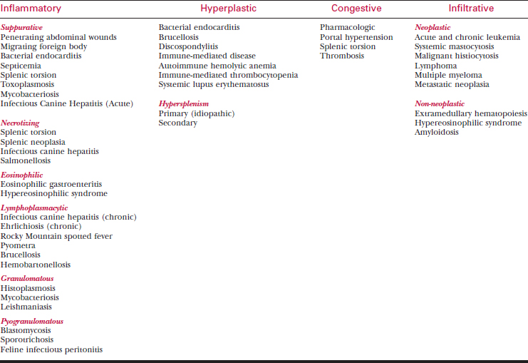
Generalized splenomegaly (Table 25-2) may result from the following:
Localized Splenomegaly
Neoplasia
• Hemangiomas also occur. Hemangiosarcoma occurs predominantly in older, large-breed dogs. German shepherds, golden retrievers, and Labrador retrievers appear to be over-represented. Most other neoplastic splenic masses are sarcomas, including fibrosarcoma, leiomyosarcoma, leiomyoma, osteosarcoma, chondrosarcoma, rhabdomyosarcoma, myxosarcoma, liposarcoma, myelolipoma, fibrous histiocytoma, lipoma, mesenchymoma, and undifferentiated sarcoma. Although lymphoma usually has an infiltrative growth pattern in the spleen, it can have a nodular appearance.
• The most common malignant neoplasms of the spleen in cats, systemic mastocytosis and lymphoma, generally have a symmetrical infiltrative pattern of growth (see below).
Nodular Hyperplasia
• Splenic nodular hyperplasia can be single or multiple nodules that are benign accumulations of lymphoid cells, hematopoietic cells, and plasma cells. The stimulus for hyperplasia may be known in some instances, such as chronic antigenic stimulation; in other cases the cause is not known.
Hematoma
• Trauma can result in subcapsular hematoma formation, causing a mass effect in the spleen. In the majority of cases, no underlying cause predisposing to intrasplenic hemorrhage is found. Splenic hematoma has been reported in association with splenic lymphoma.
• Splenic rupture associated with trauma or tumor rupture (hemangiosarcoma) may require surgical intervention.
Generalized Splenomegaly
Inflammatory and Infectious Disease
• Inflammatory changes within the spleen usually result in diffuse enlargement. A wide range of infectious diseases can result in diffuse splenomegaly. A partial list of disorders that may be associated with generalized splenomegaly is provided in Table 25-2 (see the various chapters in this book on infectious diseases for details concerning these disorders). The various types can be classified based on the primary type of cellular infiltrate (suppurative, necrotizing, eosinophilic, lymphoplasmacytic, granulomatous, or pyogranulomatous splenitis). Different etiologic agents are associated with the different cellular infiltrates.
Hyperplastic Splenomegaly
• The spleen commonly reacts to blood-borne antigens and red blood cell destruction with hyperplasia of the reticuloendothelial and lymphoid cells.
• Hyperplastic splenomegaly is common in dogs with immune-mediated disease including immune-mediated hemolytic anemia or thrombocytopenia.
• It commonly occurs in dogs with subacute bacterial endocarditis and chronic bacteremic disorders such as discospondylitis or brucellosis.
• The same occurs in certain other hemolytic disorders, including hemophagocytic syndrome, drug-induced hemolysis, pyruvate kinase deficiency anemia, phosphofructokinase deficiency anemia, familial non-spherocytic hemolysis in poodles, Heinz body hemolysis, and Mycoplasma haemofelis infection (hemobartonellosis).
Congestive Splenomegaly
• Splenic enlargement resulting from congestion can occur through a number of different mechanisms. Splenic distension occurs as a result of smooth muscle relaxation in the splenic capsule and trabeculae. Phenothiazine tranquilizers and barbiturates increase blood pooling. Pooling of blood in an enlarged spleen may account for up to 30% of the total blood volume. Consider these changes when evaluating red blood cell parameters from anesthetized or tranquilized patients.
Stay updated, free articles. Join our Telegram channel

Full access? Get Clinical Tree


