5 Diseases of the Spinal Cord
LOCALIZATION OF AND FREQUENTLY ASKED QUESTIONS ABOUT SPINAL CORD DISEASES
Yes. Animals with spinal cord disease, regardless of specific neuroanatomic location, can have variable degrees of altered muscle tone (spasticity or flaccidity), abnormalities in sensory function (ataxia, depressed or absent conscious pain perception), loss of voluntary movement (paresis/plegia), and abnormal spinal reflexes (depressed-hyporeflexia, absent-areflexia, or exaggerated-hyperreflexia).
See Tables 5-1 through 5-4.
Table 5-1 Clinical Signs of C1-C5 Spinal Cord Disease
Table 5-2 Clinical Signs of C6-T2 Spinal Cord Disease
Other signs |
Table 5-3 Clinical Signs of T3-L3 Spinal Cord Disease
Muscle tone | |
Other signs |
Table 5-4 Clinical Signs of L4-S3/Cauda Equina Disease
Muscle tone | |
Other signs |
Animals with the Schiff-Sherrington signs have opisthotonus (extension of the head and neck), spastic thoracic limbs, and paraplegia. Schiff-Sherrington posture can result from any spinal cord lesion caudal to T2.
Although the Schiff-Sherrington sign is indicative of severe spinal cord injury, it does not have any prognostic value.
Pain can be associated with disease of the paraspinal musculature, intervertebral discs, vertebral bone, meninges, spinal nerve roots, and articular facet joints.
Pain perception is transmitted by small myelinated and unmyelinated nerve fibers that are diffuse, multisynaptic, and positioned deep within the spinal cord parenchyma. These small fibers are more resistant to compressive injury than larger, more superficially located myelinated ascending nerve fibers. Thus in an animal with a transverse myelopathy, loss of deep pain implies that the damage involves a significant portion of the spinal cord diameter in that area.
CONGENITAL AND HEREDITARY ANOMALIES AND DISORDERS
Atlantoaxial Instability
AAI is a generic term used to describe any congenital or acquired disease process that allows for excessive flexion of the atlantoaxial joint. This abnormal mobility of the joint results in clinical signs consistent with a compressive cervical myelopathy due to displacement of the axis dorsally into the vertebral canal.
AAI can result from the following:
Anatomic malformations resulting in AAI are commonly seen in toy dogs or small dog breeds during the first 2 years of life. Animals with anomalous C1-C2 articulations can also be asymptomatic until experiencing what appears to be a minor traumatic incident. Traumatic lesions can affect any age, breed, or sex of dog or cat.
Clinical signs are referable to a focal C1-C5 myelopathy with severity ranging from mild cervical pain or sensory ataxia to tetraplegia with or without respiratory compromise. Evidence of dysfunction of some cranial nerves with nuclei residing in the medulla, in particular the vestibular nuclei, is present in some animals.
Radiographic indicators of AAI that can be obtained from a lateral view of the cervical spine include a widened space between the dorsal arch of C1 and the C2 spinous process and dorsocranial displacement of the body of C2 into the vertebral canal. Ventrodorsal and oblique views may reveal the anomalies or fractures of the odontoid process. The clinician should exercise extreme caution when manipulating, sedating, or anesthetizing animals with suspected AAI to prevent additional spinal cord compression. Additional radiographic techniques, such as the flexed lateral and open mouth views, have been described as being valuable diagnostic tools, but are not routinely recommended because of their potential to cause further neurologic dysfunction. Computed tomographic and magnetic resonance imaging is also beneficial in the diagnosis of AAI.
Animals with clinical signs of acute AAI should receive corticosteroid therapy (see the section on acute spinal cord trauma). Animals with clinical evidence of mild neurologic dysfunction (pain and ataxia) may be candidates for conservative management, which includes external coaptation (neck brace) and strict confinement for 4 to 6 weeks. Proper application of a neck brace is reportedly successful in some dogs. In animals with moderate to severe neurologic signs, or recurrence after conservative therapy, surgical stabilization is recommended. Several different surgical techniques using both dorsal and ventral approaches have been described.
The prognosis for animals with AAI is variable. Some positive prognostic indicators in dogs with AAI treated surgically include: age of onset of clinical signs at younger than 2 years, mild to moderate preoperative neurologic deficits, and duration of clinical signs for less than 10 months.
Dermoid Sinus
A dermoid sinus is an invagination of the skin covering the dorsum of the spinal column into the underlying tissues. It is a congenital defect that results from a faulty separation of the skin from the neural tube during development.
Dermoid sinuses have also been referred to as pilonidal sinuses and dermoid cysts.
There are five different types of dermoid sinuses. Classification is dependent on the depth to which the sinus penetrates into the underlying tissue.
Rhodesian Ridgebacks are most commonly affected, but Cocker Spaniels, Boxers, Terriers, and other breeds have been reported. In one survey of Rhodesian Ridgeback owners, 67 of 1263 (5.3%) dogs were affected with a dermoid sinus.
Signs are variable and can range from asymptomatic to severe neurologic dysfunction if the sinus communicates with the subarachnoid space. Some dogs will have clinical signs resulting from infection of the sinus and surrounding skin.
Dermoid sinuses are frequently detected during routine examinations. Dermoid sinuses occur most commonly in the cervical or craniothoracic regions, but can occur in the sacrococcygeal area and head. Multiple dermoid sinuses can occur in a single individual. In Rhodesian Ridgebacks, dermoid sinuses typically do not occur within the confines of the “ridge” of hair. Sinuses can be difficult to detect unless the haircoat is shaved, or sometimes may be palpated as a cord of tissue along the midline. Sacrococcygeal sinuses are more likely to present with dural communications. Diagnosis is further supported by plain radiography or contrast enhanced fistulography.
Treatment is often not necessary unless the sinus is infected or communicates with the dura. Surgical excision of the sinus is the treatment of choice, with some surgeons recommending a course of antibiotic treatment to resolve concurrent infection prior to excision. Cephalosporins are good empirical first choice antibiotics.
Globoid Cell Leukodystrophy
Globoid cell leukodystrophy is a lysosomal storage disease resulting from a deficiency of β-D-galactocerebrosidase, and is inherited as an autosomal recessive disorder in dogs.
Globoid cell leukodystrophy has been reported most often in the Cairn and West Highland White Terriers, but also in the Beagle and Poodle.
Pathologic findings typically include bilaterally symmetric demyelination of white matter and the perivascular accumulation of large foamy macrophages (globoid cells). White matter lesions can be of variable severity in different locations throughout the neuraxis, but typically involve the corona radiata, corpus callosum, optic tracts, peripheral subpial spinal cord parenchyma, and peripheral nerves.
Clinical signs are usually seen within the first few months of life, but can occasionally manifest after several years. Neurologic dysfunction, after it is clinically apparent, is progressive in nature. In dogs, the disease has two distinct clinical appearances. The first syndrome is dominated by a progressive upper motor neuron paraparesis. The second manifestation is primarily typified by evidence of cerebellar disease and is the most common clinical presentation in cats. Behavioral change, blindness, and hyporeflexia with muscular atrophy may also be seen in some animals. The clinical course of the disease is variable, ranging from one to several months, but is more rapidly debilitating in cats.
A specific DNA-based genetic test is available for globoid cell leukodystrophy. A clinical diagnosis can also be supported by finding globoid cells in cerebrospinal fluid (CSF), demonstrating characteristic lesions in a brain or peripheral nerve biopsy specimens, or by identifying demyelinating lesions using magnetic resonance (MR) imaging techniques. Before genetic testing, a presumptive diagnosis was often made on the basis of the appropriate signalment, history, and exclusion of other causes.
Myelodysplasia
Myelodysplasia is the nonspecific term used to refer to a multitude of spinal cord malformations that are hypothesized to be the result of failure of development or incomplete closure of the neural tube.
Individual reported defects consist of segmental spinal hypoplasia and anomalies of the central canal, including duplication, hydromyelia, syringohydromyelia, absence of a ventral median fissure, and abnormal morphologic distribution of neural elements. Several defects can occur simultaneously.
Myelodysplasia is an inherited disease in the Weimaraner and has been sporadically reported in several other breeds of dogs and cats.
Clinical signs usually suggest a thoracolumbar myelopathic process of variable severity that is first apparent at ambulation. Affected dogs may have a characteristic, symmetric “bunny-hopping” gait.
The diagnosis is made based on the signalment, history, and neurologic examination findings. Advanced diagnostic imaging modalities such as MR and computed tomography (CT) may provide further insight into the particular structural anomalies that may be present.
Currently, there is no treatment, and the prognosis is variable and dependent on the severity of the neurologic dysfunction present. The disease is not progressive in all cases, and affected animals, especially Weimaraners, may be suitable pets.
Syringohydromyelia
Syringohydromyelia is a collective term used to describe the abnormal intraspinal accumulation of fluid. Syringomyelia specifically denotes a fluid focus (i.e., syrinx) within the spinal cord parenchyma, whereas hydromyelia refers to dilatation of the central canal. Because of the difficulties associated with clinical differentiation between syringomyelia and hydromyelia, the terms are often combined and referred to as a singular entity.
Syringohydromyelia is a relatively recently recognized clinical entity in veterinary medicine, and our understanding of this condition is limited. There are several potential pathophysiologic mechanisms that may contribute to the development of syringohydromyelia. Very simplistically, these can be divided into disorders that affect normal CSF flow dynamics, resulting in altered CSF pressures, or diseases resulting in a congenital or acquired loss of normal neural parenchyma with resultant replacement with abnormal fluid accumulations.
Traumatic spinal cord injury or myelitis may result in the formation of syrinxes. Hydrocephalus and Chiari-type malformations have also been recognized in dogs with syringohydromyelia.
Chiari-type malformations are congenital or acquired abnormalities of the foramen magnum area that result in variable degrees of extension of the caudal cerebellum into the foramen magnum and caudal displacement of the medulla oblongata and fourth ventricle into the cervical spinal canal. It is the obstruction of CSF flow that results from these malformations that is thought to cause syringohydromyelia.
No; the clinical signs are related to the location of the lesion; however, although syringohydromyelia is an intraspinal disease, discomfort can be apparent. In a report of seven dogs with syringohydromyelia, cervical pain, otic pain, and thoracic limb pain were common findings. Possible sources of the pain include inflammation or hemorrhage associated with the syringohydromyelia. Dogs with syringohydromyelia may also have scoliosis or signs consistent with central cord syndrome—paresis that is more pronounced in the thoracic limbs because of selective damage to the thoracic limb pathways that are more medially located within the spinal cord. Syringohydromyelia can also be subclinical and detected as an incidental finding.
Because syringohydromyelia is an intraspinal disease, diagnosis requires visual inspection of the spinal cord parenchyma, which is ideally done with MR imaging. Occasionally, during myelography, an abnormally dilated central canal may be injected with contrast material and allow identification of syringohydromyelia. Myelography can be normal in cases in which cystic abnormalities do not communicate with the central canal and are focal, or altered CSF flow dynamics do not allow for filling of the defect.
Clinical signs have been reported to improve after the administration of antiinflammatory doses of corticosteroids or with the use of nonsteroidal antiinflammatory drugs.
The surgical management of syringohydromyelia is controversial, and will likely remain so until the pathophysiology of the condition is more thoroughly understood. Decompressive procedures involving the foramen magnum and vertebrae, cystic lesion drainage and marsupialization, and CSF fluid diversion through a variety of shunting procedures have all been attempted.
DEGENERATIVE DISEASES
Intervertebral Disc Disease
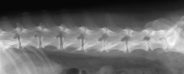
The intervertebral disc is an anatomic structure located between each pair of vertebrae of the spinal column, with the exception of C1-C2, whose physiologic role is to provide resistance against deforming loads placed on the spinal column while simultaneously allowing for some flexibility. Each intervertebral disc consists of an outer annulus fibrosus, an inner portion called the nucleus pulposus, and two cartilaginous endplates.
Type III disc disease is a clinical term used to describe an explosive herniation of a small amount of disc material that spreads a large distance within the epidural space. This type of disc disease is often associated with significant amount of epidural and subarachnoid hemorrhage, and often does not result in a large amount of spinal cord deformation. Type III disc disease is unrelated to the specific forms of intervertebral disc disease (IVDD) described by Hansen.

Figure 5-1 Lateral lumbar radiograph obtained from a mixed breed dog with paraplegia demonstrating some of the survey radiographic features of IVDD. At the L1-L2 location, the following abnormalities can be seen: wedging and narrowing of the disc space, opacification of the intervertebral foramen, and collapse of the articular facet joint.

Stay updated, free articles. Join our Telegram channel

Full access? Get Clinical Tree

