Chapter 132 Diseases of the Eyelid
Diseases of the eyelid result in a variety of clinical signs. Initially, the eyelids alone may be affected, but because of their close proximity to the cornea and conjunctiva, disease of these structures frequently results. Because corneal and conjunctival involvement is often more severe and more obvious, eyelid disease may be overlooked. Manage concurrent conjunctival and corneal disease as described in Chapters 133 and 134, respectively, in this section.
Diseases of the eyelid can be broadly categorized by their appropriate treatment (i.e., surgery or medical therapy), as listed in Table 132-1.
Surgery | Medical Treatment |
---|---|
Ankyloblepharon | Bacterial blepharitis |
Coloboma | Chalazion/hordeolum |
Entropion | Allergic blepharitis |
Ectropion | Parasitic and fungal blepharitis |
Distichiasis/ectopic cilia | |
Lagophthalmos | |
Neoplasia |
ANATOMY
• The eyelid functions to protect and moisten the cornea and to remove debris. The eyelid is covered by skin and lined internally by palpebral conjunctiva.
• The eyelid is closed by the orbicularis oculi muscle, which is innervated by the palpebral branch of the facial nerve. Paralysis of this nerve results in inability to close the eyelids. Spasm of the orbicularis oculi muscle results in spastic entropion.
• The eyelids are opened by the levator palpebrae and Mueller’s muscle, which are innervated by the oculomotor nerve and by post-ganglionic sympathetic fibers, respectively. Paralysis of either of these nerves results in ptosis, or drooping of the eyelid.
PRINCIPLES OF EYELID SURGERY
• The skin covering the eyelids is thin and easily traumatized. Perform clipping and aseptic preparation gently. A povidone-iodine solution diluted with saline solution to a 1:10 concentration is appropriate for preparing the eyelids for surgery.
• The eyelids have an extensive blood supply, and injured tissue heals well. Therefore, remove only clearly necrotic tissue. Removal of excess tissue can result in abnormal lid function.
• Close eyelid skin incisions with 4-0 to 6-0 monofilament nonabsorbable suture in a simple interrupted pattern. Close conjunctival wounds with 6-0 to 8-0 absorbable suture material. Remove nonabsorbable sutures 2 weeks following surgery.
ANKYLOBLEPHARON
Ankyloblepharon is a condition seen in neonatal puppies in which the eyelids do not open properly.
• This condition may result in a subpalpebral infection called ophthalmia neonatorum.
• If infection occurs, gently massage the eyelids to open a portion of the palpebral fissure. Occasionally, scissors or a scalpel blade is required to partially open the eyelids.
COLOBOMA
• Many cases can be corrected by everting the lid with entropion correction surgery, thus preventing corneal trauma by facial hairs.
ENTROPION
• Clinical signs vary from conjunctivitis with mild serous discharge to severe blepharospasm with corneal ulceration and purulent discharge.
• Diagnosis is made by examination of the eyelids. When the eyelid is rolled inward, facial hairs often directly contact the cornea.
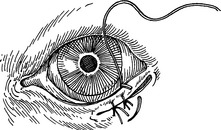
• Correct neonatal entropion in the Shar-Pei with temporary everting sutures at 3 to 5 weeks of age. If this technique fails, perform permanent surgical correction.
Classification
• Classification of entropion is made when the eyelids are relaxed. This may require topical anesthesia, palpebral nerve blocks, or general anesthesia.
Anatomic Entropion
• This may be from a defect in the eyelids themselves, or in the case of certain breeds such as the Shar-Pei and the bloodhound, from forehead or brow folds, which force the eyelids inward.
Anatomic Entropion with Secondary Spasm
• Blepharospasm exaggerates entropion such that a portion of the rolling is from an anatomic abnormality, with the remainder due to squinting.
Surgical Techniques
Ventral or Dorsal Eyelid Entropion
1. Remove an ellipse of skin parallel to and 2 to 3 mm from the lid margin (Fig. 132-1). The amount of skin removed depends on the severity of the entropion. Remove more tissue for upper lid entropion than for lower lid entropion.
2. Remove the tissue with a scalpel or by crushing the selected tissue with a hemostat and then removing the crimped tissue with scissors.
3. Close the skin routinely with monofilament nylon sutures (e.g., 4-0) in a simple interrupted pattern.
Lateral Entropion
2. If the lateral canthus is extremely lax, dissect the underlying orbicularis oculi muscle free (Fig. 132-2B) and suture it to the lateral orbital rim with 4-0 or 5-0 nonabsorbable monofilament suture material (Fig. 132-2C).