True ileal digestible lysine (g/kg)
Traditional analysis
Reactive lysine
Wheat
3.2
2.9***
Maize
2.6
1.9***
Skim milk powder
19.8
16.6***
Cottonseed meal
12.9
10.3***
Evaporated milk
23.4
20.5***
Whole milk powder
26.2
24.0***
Lactose hydrolyzed milk powder
27.2
25.1***
Popped rice cereal product
0.7
0.3***
Grain-based cereal product
1.2
0.5***
Whole grain bread
2.4
2.0**
The determination of standardised ileal amino acid digestibility coefficients for pig feeds is one of the major technical advances in pig science over the last few decades. There are economically important relative differences in determined amino acid contents between gross and digestible amino acids. However, to be able to determine ileal amino acid digestibility, digesta from the terminal small intestine (final 10–20 cm of ileum) need to be collected either in total or by sampling along with the use of a marker compound in the feed. The collection of digesta from the pig has spawned much research over the years, and there is a considerable body of knowledge on the subject. Digesta can be obtained either by dissecting the ileum from the body of the pig while under deep anaesthesia (sometimes called the “slaughter method”) or by collecting digesta from the ileo-rectal anastomised animal or from a cannulated animal. The various collection methods used in the pig have been the subject of review (Fuller 1991; Yin and McCracken 1996)
17.3 Collection of Digesta: Slaughter Method
The most straightforward way of collecting digesta from the end of the ileum is to dissect the ileum from a deeply anaesthetised animal or immediately upon euthanasia. This method is commonly used in smaller animals such as rats and chickens where the animal is killed (Payne et al. 1968; Moughan et al. 1984), and in early work with the growing pig using this approach, digesta were removed immediately after death of the animal. It is for this reason that the method is often referred to as the “slaughter method”. Because only a sample of digesta is collected with this technique, a marker compound (such as chromic oxide or titanium dioxide) needs to be included in the test diet. With the growing pig an adequate amount of digesta is usually found in the final 10–20 cm of ileum, unless a very highly digestible diet has been fed to the animal, whereupon only small quantities of digesta may be recovered. The technique has successfully been used with milk-fed piglets (Moughan et al. 1990).
There is a need, with this technique, for careful handling of the ileal tissue and attention to how the animal is killed or anaesthetised. Mucosal cells are rapidly shed into the gut lumen following death, thus increasing the protein content of the digesta. The potential problem of cell shedding can be avoided if barbiturates are used to kill the animal and digesta are collected immediately (Badawy et al. 1957; Thorpe and Tomlinson 1967), or if the ileum is removed from the anaesthetised animal.
When using the slaughter method, a frequent (usually hourly) feeding regimen may be employed, or digesta may be collected at a set time after ingestion of a single meal, to coincide with the peak digesta flow. Work has been conducted, often with the rat, studying aspects of digesta sampling (Donkoh et al. 1994a; Van Wijk et al. 1998; James et al. 2002a, b; Butts et al. 2002; Hodgkinson et al. 2002).
The advantage of this approach is its simplicity. There is minimal interference with the digestive tract prior to sampling, there are no limitations on the type of diet fed to the animal (confer cannula blockages), and it is perhaps more acceptable ethically in comparison with cannulation and anastomosis. Digesta can be sampled from several parts of the digestive tract, which is sometimes useful, but dietary markers must be used and only one terminal ileal digesta sample can be obtained per animal, so the animal cannot be used as its own control. However, the major advantage remains, that the anatomical, histological, microbial and physiological changes that can occur over time with cannulation or anastomosis are avoided with this technique.
17.4 Collection of Digesta: Ileo-Rectal Anastomosis and Cannulation Techniques
17.4.1 Ileo-Rectal Anastomosis
To avoid interference from colonic microflora and to allow repeated sampling of digesta over time, ileo-rectal anastomoses (IRA), have in the past been used to collect pre-caecal digesta from the terminal ileum. Numerous IRA configurations have been tried since they were first proposed in the early 1980s (Fuller and Livingstone 1982; Laplace et al. 1985; Picard et al. 1984). Digestibility estimates obtained using these procedures vary depending on the configuration used (Darcy-Vrillon and Laplace 1990). In all of the ileo-rectal configurations the normal functions associated with the large intestine are interrupted, and will therefore affect the absorption of water and minerals (Köhler et al. 1991). Since the first use by Fuller and Livingstone (1982) little research has been conducted to determine the effects of IRA on intestinal morphology. Salgado et al. (2002) found that anastomosed pigs had higher spleen and small intestine weights and lower large intestine weights than intact pigs. They also discovered that IRA influenced intestinal villus and crypt architecture although it had no significant effect on the activities of intestinal enzymes. Marinho et al. (2007) also found that IRA modified the intestinal villus and crypt architecture in the duodenum and ileum.
Although there are advantages of IRA there are also many disadvantages. The method has been deemed unethical by some researchers and is not permitted in some jurisdictions. Despite this the method still finds application (Branner et al. 2004; Bohmer et al. 2005; Fontes et al. 2007; Hennig et al. 2006, 2008; Hackl et al. 2010).
17.4.2 T-Cannulae
Classically, methods to obtain ileal digesta samples have involved the use of cannulae, where the lumen of the distal ileum has been exteriorised.
Cannulation techniques can be broadly divided into two categories, those where the experimental animal is fitted with a single cannula and those fitted with two, the so called re-entrant variants. Compared with single cannulation techniques, re-entrant cannulation has been thought to give more reliable nutrient digestibility values since this technique allows the collection of total ileal digesta samples within the experimental time period and this obviates the need for an indigestible marker (Yin and McCracken 1996). However, both approaches have been criticised on the grounds that such procedures have a modifying effect on the processes of digestion and absorption (Sauer and Ozimek 1986) and that differences in the determined digestibility may occur depending on the technique used (Darcy-Vrillon and Laplace 1990). Double re-entrant cannulae are rarely used today because of the complex and expensive surgical procedures.
Studies comparing the different techniques have been undertaken by a number of researchers who have all reported some disparity in the calculated digestibility coefficients (Donkoh et al. 1994b; Köhler et al. 1990; Yin and McCracken 1996; Yin et al. 2000a, b), although some of these differences may be attributable to other secondary factors including the composition of the diet, the amount of dietary fibre or the nature of the indigestible marker (Laplace et al. 1994; Sauer and Ozimek 1986; Yin et al. 2000a, b). Further discussion on the merits of these techniques is therefore required.
17.4.3 Simple “T” Cannula
First proposed by Livingstone et al. (1977a) this technique utilised a “T”-shaped cannula made of annealed Pyrex glass that had an internal diameter of just 16–20 mm (Fig. 17.1). The cannula was secured from becoming internalised by one or more Perspex washers and a self-locking nylon strap. The shank of the cannula was threaded to take an impact-resistant plastic cap. The cannula was inserted 15 cm proximal to the ileo-caecal valve. Livingstone reported that the cannulae remained intact and functional when fitted to pigs, housed in smooth-walled pens, growing to a live weight of 175 kg. Trauma from the surgery of fitting the cannulae was reported to be minimal and the intestinal activity, appetite, and growth rate of the animals was unaffected by the cannulation. They concluded that pigs fitted with such a cannula were physiologically similar to intact pigs (Livingstone et al. 1977a, b). As with all single cannula techniques, not all of the digesta flows through the cannula and the incorporation of an indigestible dietary marker to determine the total flow of digesta is essential. It is assumed that the composition of the digesta collected via the T-cannula is representative of the total quantity of digesta flowing through the distal part of the ileum, and that no fractionation of its contents, particularly between the solid and liquid phases occurs. However, this has been questioned by a number of researchers (Fuller et al. 1994; Sauer et al. 1989; Yin and McCracken 1996). Differences in the recovery of indigestible markers have indicated that phase separation may indeed occur and that this may be one reason why a greater variability in results was observed by Köhler et al. (1990) when they compared a simple T-cannula with a post-valve T-caecum cannula and a re-entrant cannula. They suggested that a reduction in the homogeneity of the samples recovered through the T-cannula may be attributable to changes in pressure at the base of the open cannula, forcing a separation of coarse and fine insoluble particles (Schröder 1988), and this might explain differences in the digestibility coefficients observed in diets rich in fibre. Although no incidence of the cannula blocking was reported by Livingstone et al. (1977a, b) this has been noted by Potkins et al. (1991), who had to change their experimental design owing to the frequency of blockages at the site of the cannula and the consequential inability to collect digesta when their experimental animals were fed a diet rich in bran fibre. Fernández et al. (2001) also reported problems with the simple T-cannulation technique, finding unreasonably large deviations in results when studying fibre rich materials. However, and despite its few disadvantages the use of simple T cannulae, for the determination of nutrient digestibilities, is one of the most commonly used cannulation techniques (Goerke et al. 2012). Today, flexible plastic and rubber T cannulae have replaced the early glass models.
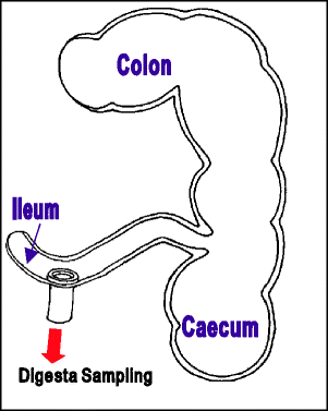

Fig. 17.1
Simple “T” cannula
17.4.4 Post-valve T-Caecum Cannula
Developed by van Leeuwen et al. (1988) in the late 1980s at the Institute for Animal Nutrition and Physiology in the Netherlands, the Post-valve T-caecum cannula (PVTC) is made of medical grade silicon tubing, with an internal diameter of 25 mm and is secured by an exterior ring and self-locking nylon straps (Fig. 17.2). The technique involves the removal of part of the caecum and installation of the cannula opposite the ileo-caecal valve. One hour before the collection of digesta starts, the cannula is opened to allow the ileo-caecal valve to protrude into the lumen of the cannula. Although this ensures that more of the digesta flowing from the ileum is collected, this technique cannot guarantee a complete quantitative collection of ileal digesta and so the inclusion of an indigestible dietary marker is advised (van Leeuwen et al. 1991). Having studied more than 100 animals fitted with PVTC cannulae, with live weights ranging from 8 to 100 kg, van Leeuwen’s group found that post-operative recovery was swift and that the animals displayed no signs of discomfort or a reduction in appetite, even over long periods of time. Following the partial caecectomy and insertion of the cannula into what remains of the caecum, the physiology of both the ileum and the colon appear to be normal. As the gut is not transected, there appears to be no interference with the myoelectric innervation of the intestines that may reduce intestinal motility, points that have been corroborated by a number of other researchers (Köhler et al. 1990, 1992a, 1992b; Yin and McCracken 1996; Yin et al. 2000b). Although few digestibility studies have compared PVTC cannulae with other methods of collection, those that have concluded that results, using the PVTC technique, are comparable with those obtained using simple T-cannula (Köhler et al. 1990; Yin and McCracken 1996; Yin et al. 2000b).
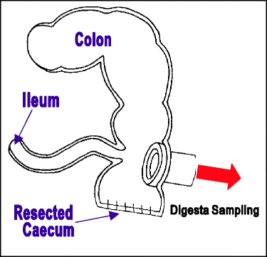

Fig. 17.2
Post-valve T-caecum cannula
In one long-term study by Kohler et al. (1992a, b) using 23 pigs (9 with IRAs, 7 with PVTCs and 6 intact pigs), no differences were found in growth performance, body nitrogen retention, blood variables and mineral balances, between intact animals and those fitted with PVTCs.
Greater precision can be achieved using the PVTC method for most diets and this is thought to be attributable to greater consistency in the recovery of the indigestible marker, specifically titanium dioxide. However, with high fibre diets some variability in the amount of digesta recovered has been recorded, which confirms the necessity of using an indigestible marker (Köhler et al. 1990, 1991; van Leeuwen et al. 1991; Yin et al. 2000a) Although the PVTC cannulation technique has been found to have a minor effect on total tract nutrient digestibility (Lindberg 1997), it is at present considered to give the most satisfactory results for ileal digestibility determination (Hodgkinson and Moughan 2000a; Presto et al. 2011).
17.4.5 Steered Ileo-Caecal Valve
Mroz et al. (1994, 1996) developed a variant of the PVTC technique which they termed the steered ileo-caecal valve (SICV), cannulation (Fig. 17.3). This technique permits the collection of digesta, with both low and high fibre diets, and maintains the normal physiological function of the gut as it does not require the removal or transection of any part of the GIT. Although this technique was thought to allow the quantitative collection of digesta, the recovery of a chromium marker from the faeces of cannulated pigs was approximately 20 % less than in intact pigs. Therefore under the conditions of their study, the use of an inert marker was deemed advisable for determination of the total tract digestibility and ileal digestibility of nutrients. Changes proposed by Radcliffe et al. (2005) resulted in a decrease in post-surgical complications, notably fewer and less severe adhesions, together with less leakage of digesta around the cannulae. The surgery for this technique is more complex than the PVTC method, but it appears to be less prone to blockages.
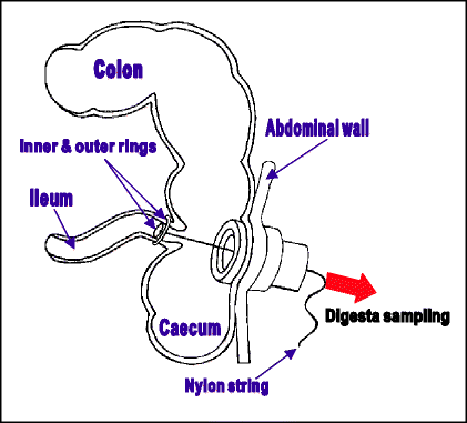

Fig. 17.3
Steered ileo-caecal valve
In comparison to the simple T cannula, Zhang et al. (2004) concluded that ileal digesta samples collected using the SICV-cannula were more homogenous.
17.4.6 Simple Ileo-Ileal Re-entrant Cannula
Although re-entrant fistulae have been commonly used to study the digestive process in ruminants (Phillipson 1952), Cunningham et al. (1962, 1963) used a similar re-entrant cannula to compare the digestibility and rate of passage of a variety of purified and natural feed materials in the gut of the pig (Fig. 17.4). Blockages would often occur in the cannulae, particularly, if coarsely ground feeds were administered, but the cannulae were well tolerated by the pigs. Kowalik et al. (2004) studying the digestion of starch and crude fibre in segments of the digestive tract of sheep reported no problems with blockages.
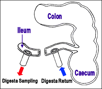

Fig. 17.4
Simple ileo-ileal re-entrant cannula
17.4.7 Ileo-Caecal Cannula
Both Cunningham et al. (1963) and later Cho et al. (1971) found that the flow of digesta through the externally joined ileo-ileal re-entrant cannulae was not uniform. Easter et al. (1972) and Easter and Tanksley (1973) believed that the surgically disrupted small intestine was not capable of forcing digesta through the externalised cannulae and the ileo-caecal valve. As the ileo-caecal valve was not essential to the digestion process (Davenport 1966), changing the configuration of the re-entrant cannula from ileo-ileal to ileo-caecal offered a solution to this resistance (Fig. 17.5). Although the use of single caecal cannulae in the digestion studies of pigs had been demonstrated by earlier researchers (Henderickx et al. 1964; Redman et al. 1964), this was the first time that a double, ileo-caecal re-entrant had been used. Although not widely used, ileo-caecal re-entrant cannulae are still employed particularly in studies involving dietary fibre (Bartelt et al. 1999; Rapp et al. 2001).
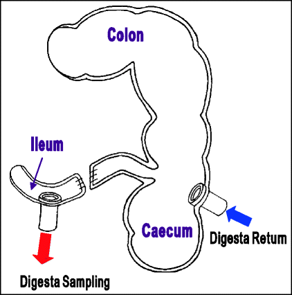

Fig. 17.5
Ileo-caecal cannula
17.4.8 Ileo-Colic Post-valve Fistulation
First described by Darcy et al. (1980), Ileo-colic post-valve fistulation (ICPV), which they considered to be a reference method (Fig. 17.6), is surgically difficult and too time consuming for routine ileal digestibility measurement. Early suggestions that this method could be used in diets rich in fibre (Sauer and Ozimek 1986) were recanted by Fuller et al. (1994) who reported evidence to the contrary for diets rich in barley.
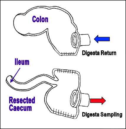

Fig. 17.6
Ileo-colic post-valve fistulation
17.4.9 Comparative Summary of Ileal Digesta Collection Techniques
There is little agreement between researchers as to which digesta collection technique is the best, although lately the PVTC method has been popular. Variations in determined ileal amino acid digestibility values have been noted due to differences in:
Indeed Fuller et al. (1994), state that each method has its strengths and limitations such that no single method is suited for all purposes, a point echoed more recently by Knudsen et al. (2006) when reviewing in vivo methods of studying the digestion of starch in pigs and poultry. Such variation is also compounded by the differing performance of indigestible markers necessary to quantify the total flow of digesta (Yin et al. 2000b). A comparative summary of the advantages and disadvantages of the various collection techniques is given in Tables 17.2 and 17.3.
Table 17.2


Advantages and disadvantages of different cannulation techniques
Stay updated, free articles. Join our Telegram channel

Full access? Get Clinical Tree


