Chapter 64 Dentistry and Diseases of the Oropharynx
Dental disease and diseases of the oropharynx are common in dogs and cats. These diseases can be divided into categories including oral surgical disease, periodontal disease, endodontic disease, orthodontic disease, stomatitis/gingivitis, neoplastic disease, and salivary gland disease. Neoplastic disease of the maxilla and mandible is discussed in Chapter 99.
ORAL SURGICAL DISEASE
The two major categories of oral surgical (nonneoplastic) disease are:
Anatomy
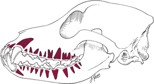
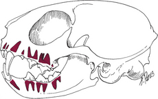
Table 64-2 TOOTH ROOTS IN DOGS AND CATS
| Type of Tooth | No. of Roots |
|---|---|
| In Dogs | |
| Incisor | 1 |
| Canine | 1 |
| Maxillary (upper) cheek teeth: | |
| 1st (1st P) | 1 |
| 2nd and 3rd (2nd and 3rd P) | 2 |
| 4th–6th (4th P; 1st and 2nd M) | 3 |
| Mandibular (lower) cheek teeth: | |
| 1st and last (1st P; 3rd M) | 1 |
| 2nd–6th (2nd–4th P; 1st and 2nd M) | 2 |
| In Cats | |
| Incisor | 1 |
| Canine | 1 |
| Maxillary (upper) cheek teeth: | |
| 1st (2nd P) | 1 |
| 3rd (4th P) | 3 |
| All others | 2 |
| Mandibular (lower) cheek teeth: | |
| All | 2 |
M, molar; P, premolar.
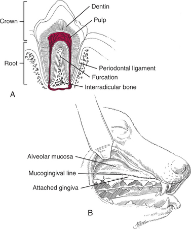
Table 64-3 BASIC ANATOMIC STRUCTURES OF THE DENTITION
| Crown: | Portion of the tooth located in the oral cavity. It is covered by enamel. |
| Root: | Portion of the tooth that lies within the alveolar bone. It is covered by cementum. |
| Furcation: | The space between the roots of a multirooted tooth. |
| Periodontal ligament: | Ligament that attaches the root of the tooth to the alveolar bone. |
| Interradicular bone: | Bone located between the roots. |
| Buccal bone: | Bone located on the buccal, or cheek, side of the tooth root. |
| Attached gingiva: | Keratinized gingiva firmly attached to the underlying alveolar bone. |
| Alveolar mucosa: | Mucosa that is loosely attached to the underlying bone. |
| Mucogingival line: | Anatomic landmark that separates the attached gingiva from the alveolar mucosa. |
Diseases Requiring Dental Extraction
Diagnosis and Indications for Extraction
The decision to perform dental extraction depends not only on the dental disease present but also on the client’s ability and desire to pursue alternatives to extraction, such as periodontal, endodontic, and orthodontic therapy.
Gross Decay/Dental Erosion
Periapical Inflammation
Impacted Teeth
Impacted (unerupted) teeth may cause nasal discharge, orthodontic problems, and bone loss.
Deformed Teeth
Preoperative Considerations
Surgical Procedure
Equipment
Table 64-4 DENTAL EQUIPMENT APPLICATIONS
| Equipment | Applications |
|---|---|
| Portable electric dental drill with low-speed hand piece and prophy angle (Vetroson Millennium Motor Pack, Henry Schein, Inc.; Port Washington, NY) | Sectioning teeth, removal of alveolar bone, polishing teeth |
| Mobile delivery system with high-speed handpiece, low-speed handpiece, water and air syringe (Vet-Base, Henry Schein, Inc.) | Sectioning teeth, removal of alveolar bone, polishing teeth, cavity and crown preparations |
| Piezoelectric scaler, electric or air driven (Spartan USA, Inc.; Fenton, MO) | Permits rapid, easy removal of dental calculus |
| Dental radiography unit (AFP Imaging, Elmsford, NY) | Permits rapid, easy exposure of dental radiographs |
Table 64-5 DENTAL INSTRUMENTATION
| Instrumentation* | Applications |
|---|---|
| Oral Surgery | |
| Dental elevator | Tears periodontal ligament during extraction |
| Dental luxators | Has a thinner, sharper blade than traditional elevators |
| Extraction forceps | Completes breakdown of periodontal ligament and removal of tooth from alveolus during extraction |
| Root tip pick | Removal of small-breed deciduous teeth or broken root tips |
| Bone curette | Debridement of alveolus following extraction |
| Periosteal elevator | Elevation of mucoperiosteum during oronasal fistula repair and palatal surgery |
| Periodontic | |
| Periodontal probe/explorer | Measurement of depth of periodontal pocket (probe); detection of pulpal exposures and carious lesions (explorer) |
| Hand scalers | Subgingival scaling |
| Curette (Columbia #13/14) | Removal of accretions on the root surface and removal of granulation tissue from the pocket wall |
| Prophylaxis paste | Polishing teeth following scaling |
| Endodontic | |
| Endodontic files (Hedstrom/K-Files) | Debridement of necrotic pulpal tissue from canal |
| Root canal plugger | Compression of gutta percha into the apex |
| Light-Speed and SimpliFil System† | |
* Available from Henry Schein, Inc.; Port Washington, NY and from Cislak Manufacturing Inc, Niles, IL.
† Available from Light Speed Endodontics, San Antonio, TX.
| Material | Applications |
|---|---|
| Composite resins (Prodigy, sdsKerr, Orange, CA) | Aesthetic restorations following endodontic therapy or cavity preparation |
| Glass ionomers (Vitrebond, 3M EPSE, St. Paul, MN) | Restoration of feline external root resorptive lesions |
| Endodontic sealers (AH Plus, Dentsply, Konstanz, Germany) | Endodontic filling material |
| Mineral trioxide aggregate (ProRoot MTA, Dentsply Tulsa Dental, Tulsa, OK) | Stimulates closure of an apex and reparative dentin formation following spulpotomies |
| Gutta percha (Successfil, Hygenic) | Fills the pulp canal following debridement during root canal therapy |
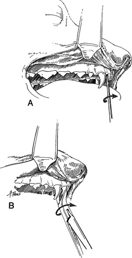
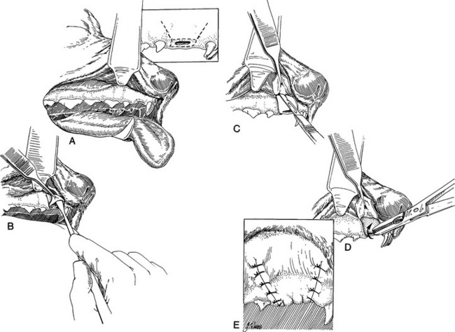
Simple Extraction
Simple extraction refers to the extraction of a small single-rooted tooth such as an incisor.
Technique
Multirooted Extraction
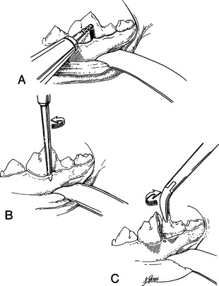
Complicated Surgical Extraction
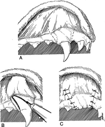
Technique
Postoperative Care and Complications
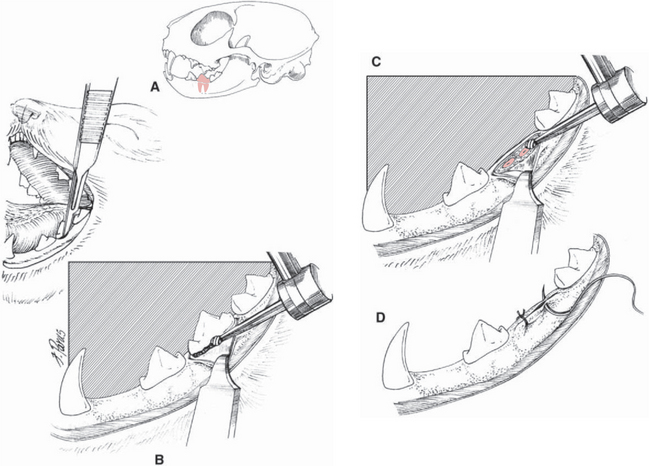
Broken Root Tips
Oronasal Fistulas and Palatal Defects
Anatomy
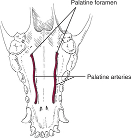
The surgical anatomy varies with the location of the defect. The mucoperiosteal flap used in the repair of oronasal fistulas extends from the margins of the defect across the mucogingival line (the anatomic landmark separating the attached gingiva from the alveolar mucosa). Surgical techniques used to repair defects in the hard palate often require preservation of the palatine arteries, which arise approximately 1 cm palatal to the maxillary fourth premolar and course rostrally along the hard palate (Fig. 64-9).
Etiology
Palatal defects may be congenital or acquired.
Acquired Defects
Clinical Signs
Diagnosis
Preoperative Considerations
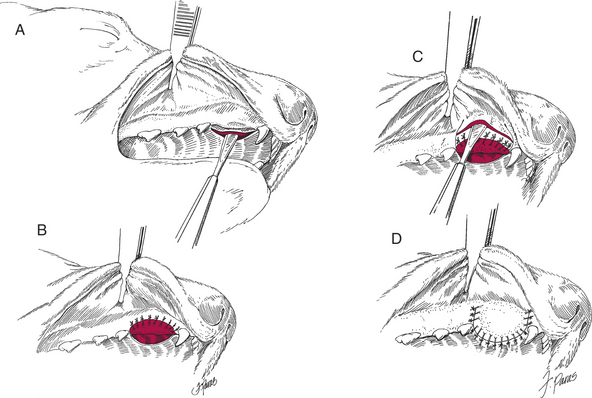
Surgical Procedure
Single-Layer Mucoperiosteal Flap
Technique
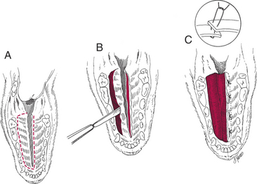
Double-Layer Mucoperiosteal Flap
Technique
Overlapping Flap Technique
Technique
Postoperative Care and Complications
Stay updated, free articles. Join our Telegram channel

Full access? Get Clinical Tree


