Chapter 3 Dental Radiology
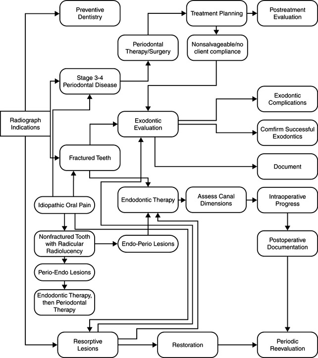
In the United States during the 1970s, dental radiology was not incorporated routinely in the course of extractions and other procedures. During the 1980s, practitioners who were interested in performing at a more advanced level found that radiographs helped them. At first, the convenient method was to use the larger veterinary medical units in the office. As radiology was recognized as more useful, these enterprising veterinarians explored the practicality of purchasing human dental radiographic units. It quickly became apparent that a definitive diagnosis often was not made unless intraoral radiographs were taken. Dental radiography has now become an essential part of the veterinary dental diagnostic workup. The diagnostic yield of full mouth radiographs in feline and canine patients is high, and routine full mouth radiography is justified.1,2 These studies found that if disease existed, radiographs were clinically useful in 86.1% of the cases in the study (Tables 3-1 and 3-2). Follow-up radiographs may be required, and professional judgment should be used in determining the type, frequency, and extent of each radiographic examination.1
Table 3-1 VALUE OF RADIOGRAPHS—NO CLINICAL FINDINGS PRESENT
| Dogs | Cats | |
|---|---|---|
| Incidental radiographic findings | 41.7% | 4.8% |
| Clinically important findings | 27.8% | 41.7% |
| Radiographs of no value | 30.5% | 53.6% |
Table 3-2 VALUE OF RADIOGRAPHS—CLINICAL FINDINGS PRESENT
| Dogs | Cats | |
|---|---|---|
| Confirmational only | 24.3% | 13.9% |
| Additional findings | 50.0% | 53.9% |
| Clinically essential findings | 22.6% | 32.2% |
| No value | 3.1% | 0 |
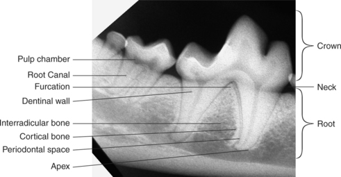
Normal Radiographic Anatomy1–5
Normal and Abnormal
Reading the Radiograph
RADIOGRAPHIC FINDINGS
Structures Visible
Normal Young Patient
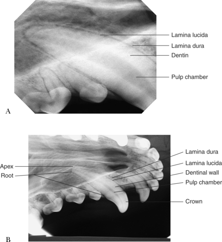
Normal Older Animal
To summarize, the normal radiographic signs of aging are as listed below:
Indications
Contraindications
Radiographic Units
Medical Radiographic Unit
Dental Radiographic Unit
Table 3-3 60 kV (MAXIMUM IMAGE CONTRAST)
| Small dog or cat | Large dog | |
|---|---|---|
| Incisor | 0.25 second | 0.4 second |
| Canine | 0.25 second | 0.4 second |
| Premolar | 0.32 second | 0.5 second |
| Lower molar | 0.4 second | 0.63 second |
| Upper molar | 0.5 second | 0.8 second |
Table 3-4 70 kV (MAXIMUM GREY LEVEL DEFINITION)
| Small dog or cat | Large dog | |
|---|---|---|
| Incisor | 0.125 second | 0.2 second |
| Canine | 0.16 second | 0.25 second |
| Premolar | 0.2 second | 0.32 second |
| Lower molar | 0.25 second | 0.4 second |
| Upper molar | 0.32 second | 0.5 second |
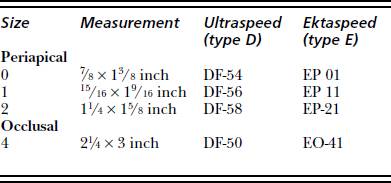
Materials
Film
Film Identification
Identification Methods
Identifying Right and Left Sides
Film Storage
TAKING AN INTRAORAL RADIOGRAPH4,5,7–10
General Comments
Whole Mouth Survey: Small Dog or Cat9
Whole Mouth Survey: Large Dog9
Stay updated, free articles. Join our Telegram channel

Full access? Get Clinical Tree


