42 CYTOLOGY OF NEOPLASIA
1 What is a good general approach to evaluation and classification of tissue cells in cytologic preparations?
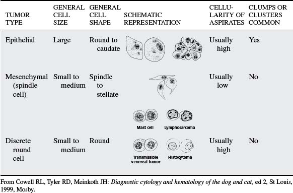
2 Discuss the cytologic criteria of malignancy.
In general, a uniform population of cells suggests the mass is benign, whereas variation in cells suggests malignancy. An important exception is lymphoma (lymphosarcoma), which consists of a fairly uniform population of lymphoblasts, whereas lymphoid hyperplasia typically has marked variation due to the mixed population of small lymphocytes, prolymphocytes, lymphoblasts, and plasma cells. Nuclear criteria of malignancy are considered diagnostic, whereas cytoplasmic criteria of malignancy are only supportive of malignant neoplasia. It is important to find more than three of the following nuclear criteria of malignancy in a few cells to many cells to call a mass “malignant neoplasia” (Table 42-2).
Table 42-2 Criteria of Malignancy in Tissue Cells
| CRITERIA | DESCRIPTION | SCHEMATIC REPRESENTATION |
|---|---|---|
| General Criteria | ||
| Anisocytosis and macrocytosis | Variation in cell size, with some cells ≥1.5 times larger than normal |  |
| Hypercellularity | Increased cell exfoliation due to decreased cell adherence | Not depicted |
| Pleomorphism (except in lymphoid tissue} | Variable size and shape in cells of the same type | 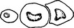 |
| Nuclear Criteria | ||
| Macrokaryosis | Increased nuclear size |  |
| Increased nuclear/cytoplasm (N/C) ratio | Normal nonlymphoid cells usually have N/C of 1:3-1:8 depending on the tissue; increased ratios (e.g., 1:2, 1:1) suggest malignancy. | See Macrokaryosis. |
| Anisokaryosis | Variation in nuclear size; especially important if nuclei of multinucleated cells vary in size |  |
| Multinucleation | Multiple nuclei in a cell; especially important if nuclei vary in size |  |
| Increased mitotic figures | Mitosis is rare in normal tissue. | 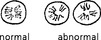 |
| Abnormal mitosis | Improper alignment of chromosomes | See Increased mitotic figures. |
| Coarse chromatin pattern | Chromatic pattern is coarser than normal and may appear ropy or cordlike. |  |
| Nuclear molding | Deformation of nuclei by other nuclei within same cell or adjacent cells | 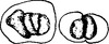 |
| Macronucleoli |  | |
| Angular nucleoli | Nucleoli are fusiform or have other angular shapes instead of normal round to oval shape. |  |
| Anisonucleoliosis | Variation in nucleolar shape or size; especially important if variation is within same nucleus. | See Angular nucleoli. |
Modified from Cowell RL, Tyler RD, Meinkoth JH: Diagnostic cytology and hematology of the dog and cat, ed 2, St Louis, 1999, Mosby.
Nuclear Criteria of Malignancy
Stay updated, free articles. Join our Telegram channel

Full access? Get Clinical Tree


