43 CYTOLOGY OF LYMPH NODES AND THYMUS
3 What is the cytologic appearance of an aspirate from a normal lymph node?
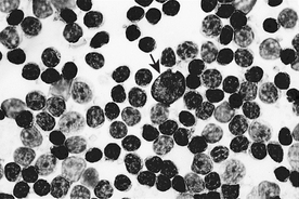
Aspirates from normal lymph nodes consist of 75% to 95% (usually about 90%) small mature lymphocytes (Figure 43-1). Small lymphocytes are slightly larger than the size of a canine red blood cell (RBC) and about 1.5 to 2 times the size of a feline RBC. They have a dark staining, roundish nucleus with densely aggregated chromatin and no visible nucleoli. Small lymphocytes have a scanty amount of clear to light-blue cytoplasm that is generally visible only on one side of the nucleus (not visible all the way around nucleus). Other cell types are present in low numbers and may include lymphoblasts, prolymphocytes, plasma cells, macrophages, neutrophils, and mast cells. Lymphoglandular bodies, bare nuclei, and strands of nuclear chromatin are often present as well.
5 What is the cytologic appearance of a hyperplastic lymph node, and how does it differ from a normal lymph node?
7 What is a Mott cell?
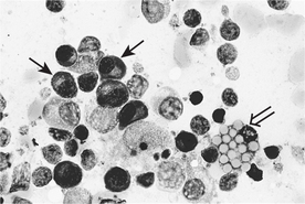
A Mott cell is a plasma cell that has its cytoplasm filled with roundish vacuoles that stain clear to light blue (Figure 43-2). These vacuoles, termed Russell bodies, are packets of immunoglobulin.
Stay updated, free articles. Join our Telegram channel

Full access? Get Clinical Tree


