Figure 51.2 Transabdominal ultrasound image of the urinary bladder in an alpaca. The ideal site for needle insertion is the curvature of the urinary bladder as it contacts the body wall.
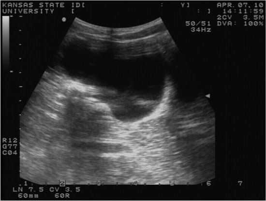
Figure 51.3 Use of an 18-gauge spinal needle, with concurrent ultrasound guidance, to perform cystocentesis.

Table 51.1 Urinalysis reference ranges for llamas at sea level (Adapted from Fowler, 1998).
| Color | Clear, light yellow-amber | |
| Specific Gravity | 1.013–1.048 (mean 1.023) | |
| pH | 7–8.5 | |
| Protein | Negative | Some 1+ |
| Glucose | Negative | Some 1−3+ |
| Ketones | Negative | Some 1+ |
| Urobilinogen | Negative | Some 2−3+ |
| Bilirubin | Negative | |
| Blood | Negative | |
| Sediments | Calcium Oxalate common | |
| Uric Acid rare | ||
POTENTIAL COMPLICATIONS
The procedure is contraindicated in patients with a small volume of urine in the urinary bladder, obstruction of urine outflow from the bladder if surgery is not planned, recent abdominal surgery, suspected bleeding disorders or caudal abdominal tumors. Risks include urinary bladder laceration, rupture, laceration of bowel with subsequent peritonitis. In general, cystocentesis is discouraged in camelids. The urinary bladder is intra-pelvic in location unless distended, and thus, risk of bowel perforation is significant. Ultrasound-guided cystocentesis is always preferred. In cases of urinary outflow obstruction, bladder pressure is sufficient to cause persistent leakage after cystocentesis in some camelids. Thus, this procedure is not suited for transient alleviation of bladder distension unless surgery or other means of clearing the urinary obstruction are performed soon afterward.
PATIENT MONITORING/AFTERCARE
The patient should be monitored for normal urination and the development of uroperitoneum. Caution should be exercised for patients with bacterial cystitis or other urinary bladder wall compromise because of increased risk of urine leakage and septic peritonitis.
RECOMMENDED READING
Ballweber LR. 2009. Ecto- and endoparasites of New World Camelids. Vet Clin N Am Food Anim Pract; 25(2):295–310.
Fowler ME. 1998. Medicine and Surgery of South American Camelids, 2nd ed. Ames:Blackwell.
Lackey MN, Belknap EB, Salman MD, et al. 1995. Urinary indices in llamas fed different diets. Am J Vet Res; 56(7):859–865.
McLaughlin BG, Evans NC. 1989. Urethral obstruction in a male llama. J Am Vet Med Assoc; 195(11):1601–1602.
Miesner MD. 2009. The straining camelid: GI or urinary? In: Proceedings of the 81st Western Veterinary Conference 2009. Available at: http://wvc.omnibooksonline.com/data/papers/2009_V559.pdf
Stay updated, free articles. Join our Telegram channel

Full access? Get Clinical Tree


