Procedure
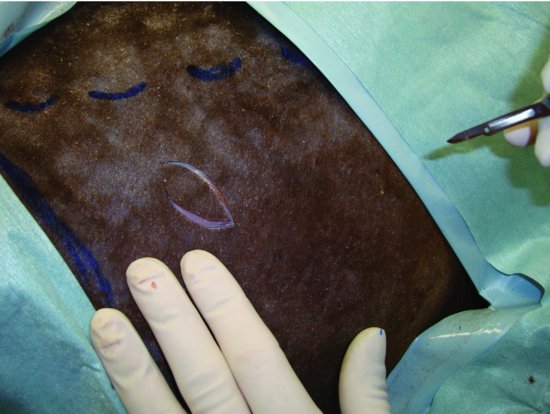
The incision for this procedure is centered in the lateral abdominal wall approximately 6- to 8-cm caudal to the last rib and 6- to 8-cm ventral to the transverse processes of the lumbar vertebrae. For temporary C1 stoma, a linear skin incision is made perpendicular to the spine. For permanent or long-term use C1 stomas, an elliptical skin incision is made approximately 3 cm in length and 2 cm in width (Figure 45.2). Larger stomas are possible, and permanent C1 cannulas may be placed in camelids (using commercially available cannulas designed for use in goats), but these are associated with greater morbidity and risk of death. The incision is continued through the muscles of the lateral abdominal wall using Metzenbaum scissors by separating the muscle parallel to the direction of the muscle (external abdominal oblique cranial-dorsal to caudal-ventral; internal abdominal oblique caudal-dorsal to cranial-ventral; transversus abdominus dorsal to ventral). (See Figure 45.3). The peritoneum is grasped using Brown-Adson forceps and penetrated using Metzenbaum scissors and the dorsal-lateral aspect of the C1 grasped using Allis tissue forceps or a penetrating towel clamp and exteriorized from the abdomen. Stay sutures (e.g., #0 PDS or PGA-910) are placed through the external abdominal oblique muscle and seromuscular layer of the C1 at 4 equidistant places along the circumference of the stoma site (e.g., using a clock-face template, these sutures would be placed at the 12, 3, 6, and 9 o’clock positions; Figure 45.4 and 45.5). These stay sutures help to protect the stoma suture from tension. The C1 stoma is made using simple continuous sutures placed through the skin and seromuscular layer of C1 (Figure 45.6). The simple continuous patterns are interrupted at 120-degree arcs around the circumference of the stoma (e.g., using a clock-face template, three segments of simple continuous suture would be done from 12 to 4 o’clock, 4 to 8 o’clock, and 8 to 12 o’clock). (See Figure 45.7.) Interruption of the simple continuous suture lines prevents formation of a purse string and allows better control of the tension along the suture lines. Finally, a vertical incision is made into the lumen of C1 being careful not to compromise any of the prior placed sutures (Figure 45.8). These stomas are expected to heal closed within 4 to 6 weeks unless maintained open by use of indwelling cannulas. If the stoma does not heal closed, these stomas can be surgically reconstructed by en bloc resection of the stoma and careful reconstruction of each tissue layer. Surgical reconstruction is most easily done with the patient under general anesthesia. For permanent stomas, the C1 stoma can be made first and then the cut edge of the C1 sutured to the cut edge of the skin in such a way that these tissues heal together to form a permanent fistula from the skin to C1. These procedures require careful attention to detail to prevent contamination of the abdomen.
Stay updated, free articles. Join our Telegram channel

Full access? Get Clinical Tree


