EQUIPMENT NEEDED
The following equipment is needed: eyelid speculum, Steven’s tenotomy scissors, Colibri or tying forceps, ophthalmic needle holders, 6-0 to 8-0 polyglactin 910 suture (smaller is better), and magnifying lens (Gelatt, 2007).
RESTRAINT/POSITION
General anesthesia is used.
TECHNICAL DESCRIPTION OF PROCEDURE/METHOD
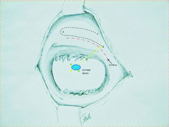
This technique has been described by multiple people in multiple species. This technical description was made using Gellat’s Veterinary Ophthalmology text as a reference and with personal consultation with Dr. Anne Metzler, DVM, MS, ACVO. Specific location and size of flap is determined by the corneal defect being covered. The conjunctiva is incised at a position that will allow undermining and transposition to the corneal lesion. The initial incision should be perpendicular to the cornea and undermined parallel to the corneal edge with more width than the abnormal corneal tissue to allow for some contracture (Figure 72.2). Careful dissection is done to remove the conjunctiva from Tenon’s capsule without putting holes in the conjunctiva. The pedicle is created by making two long incisions in the undermined conjunctiva at and parallel to the limbus and at a parallel line distal to the limbus creating a flap that is 1–2 mm wider than the defect to be covered and wider at the base than at the tip (Figure 72.2). This creates a pedicle that is roughly rectangular in shape with three sides cut free and one edge attached. Final graft material should be thin enough that the scissors can be seen through it, large enough to more than cover the defect, loose enough to have no tension when sutured in place, and the base should be wider than the tip of the graft.
Figure 72.2 Schematic drawing of camelid eye with corneal lesion and conjunctival flap harvest. Initial incision is perpendicular to the cornea at A and undermined toward B and C. The location of A is determined by measuring from a location nearest to the lesion, which will be the base of the pedicle at B to 1–2 mm beyond the lesions distal edge (light green dotted line). The width of the flap is determined by the size of the lesion (dark green dotted line). The base of the pedicle must be wider than the tip. After elevation of the conjunctiva, the pedicle is severed from A to B along the limbus, then from A to C. (Illustration courtesy of Matt Miesner.)

Stay updated, free articles. Join our Telegram channel

Full access? Get Clinical Tree


