Computed Tomography
Musculoskeletal
Basic Information 
Overview and Goal(s)
• Computed tomography (CT) is most commonly used in equine practice for osseous studies (eg, complex fractures, sequestrum identification, the skull), but recent work has documented the utility of CT for soft tissue evaluation.
• The relatively small size of the CT opening (gantry or bore) limits usefulness to the extremities, from the carpus and tarsus distally, the skull, and proximal to mid-cervical spine, in most adult horses. In addition, special tables are required for adult horses because the limit for most CT scanners is around 400 lb.
• CT uses a rotating x-ray tube to produce axial (transverse or cross-sectional) tomographic images of the patient as the CT table advances in predetermined small distances after each slice of data acquisition. These axial images are organized into spatially consecutive, parallel image sections (tomographic slices).
• Reconstruction of axial CT images into sagittal or dorsal image planes is possible. Reconstructions into any plane necessary for image analysis are also possible, including three-dimensional images. This is unlike magnetic resonance imaging (MRI), in which each image plane must be acquired separately, increasing the time of MRI studies relative to CT.
• In CT, a computer stores x-ray attenuation data and generates a matrix of picture elements termed pixels. The x-ray attenuation data of each pixel are represented as a shade of gray.
• Each shade of gray is assigned a CT number in Hounsfield units (HU). In this scale, water is equal to 0 HU, pure air is equal to −1000 HU, and cortical bone is 1000 HU or greater. The density level of almost all soft tissue organs is between 10 and 90 HUs.
• The viewed CT image is thus composed of a series of pixels forming the matrix; the matrix is a square series of pixels forming the field of view (FOV). Typical matrix size for CT scanners is 512 × 512 pixels, or more than 260,000 pixels comprising the image.
• Because CT acquires tomographic slices, each pixel actually represents a small volume of tissue, a voxel. For example, a 1 × 1 mm pixel may represent a 1 × 1 × 10 mm volume of tissue if the tomographic slice is 10 mm thick. It is important to remember that matrix size and slice thickness are responsible in large part for image resolution; each pixel (and therefore voxel) can only represent one particular HU value (only one shade of gray).
• There are two types of image resolution that are important: spatial resolution and contrast resolution. Spatial resolution is the ability to differentiate small anatomic structures from one another. CT excels in spatial resolution. Conversely, MRI has much better tissue contrast resolution than CT. This is why MRI has become the standard for orthopedic injuries and neurologic disease in human and veterinary medicine.
• CT is capable of differentiating 3000 to 4000 levels of HUs. The viewing monitor can display a maximum of 256 gray tones at one time, whereas the human eye is able to discriminate only about 20.
• CT displays of various tissues and regional anatomy are therefore selected and enhanced by operator-adjustable viewing parameters, termed window level and window width. Adjusting the gray scale is a feature of CT that allows viewing a large range of tissues from a single acquisition series. This is unlike MRI, in which multiple sequences must be obtained to view various tissues and tissue characteristics. This is one reason why CT studies typically require much less time to acquire than MRI studies.
• The window level (L) is the center point of HU value display. Window level should be set close to the mean density level of the tissue to be critically examined. The window width (W) is a range of HU values displayed and influences the contrast of the image.
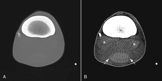
• Commonly used terminology includes “bone window” when window and level are set to maximize bone detail and “soft tissue” window when parameters are set to maximize soft tissue differentiation (Figure 1).
Equipment, Anesthesia
• The patient is typically positioned in lateral recumbency, usually with the limb of interest dependent to minimize respiratory motion.
• Initial pilot images (“scout films”) are made to center the limb and position it perpendicular to the plane of gantry/table translation.
Stay updated, free articles. Join our Telegram channel

Full access? Get Clinical Tree


