Common Protozoans That Infect Domestic Animals
Learning Objectives
After studying this chapter, the reader should be able to do the following:
Key Terms
Flagellates
Amoebae
Ciliates
Apicomplexans
Trophozoite
Cyst
Endosome
Hemoprotozoan
Phlebotomine sand flies
Canine piroplasm
Ring form
Gamonts
Commensals or commensalism
Equine protozoan myeloencephalitis (EPM)
Infectious enterohepatitis or blackhead
Ichthyophthiriosis
This chapter is a continuation of Chapter 10 in that it details the common protozoans that infect domestic animals. The parasitic protozoans are discussed in the same order as in Chapter 10: flagellates, amoebae, ciliates, and apicomplexans. These parasites are discussed in the same system-by-system manner, that is, those infecting the gastrointestinal tract, the circulatory system and peripheral blood, and the urogenital system. These parasites are discussed in the following species of domesticated animals: dogs and cats, ruminants (i.e., cattle and sheep), horses, pigs, pet and aviary birds, domestic fowl, rabbits, mice, rats, hamsters, guinea pigs, and fish.
Dogs and Cats
Gastrointestinal Tract
Protozoans that infect the gastrointestinal tract of dogs and cats are flagellates, amoebae, ciliates, and apicomplexans.
Flagellates
Parasite: Giardia species
Host: Dogs, cats, horses, ruminants, and exotic species
Location: Intestinal mucosa
Distribution: Worldwide
Derivation of Genus: Named after a famous protozoologist, Giard
Transmission Route: Ingestion of oocysts
Common Name: Giardia
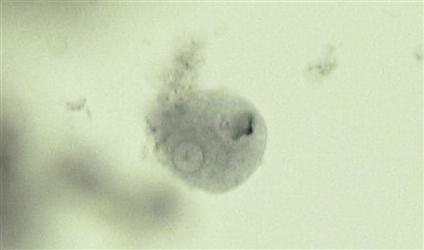
Giardia species are flagellated protozoans often recovered from the feces of dogs and cats with diarrhea. They may also be recovered from animals with normal stools. These parasites occur in two morphologic forms: a rarely observed, motile feeding stage (the trophozoite) and a frequently observed, resistant cyst stage.
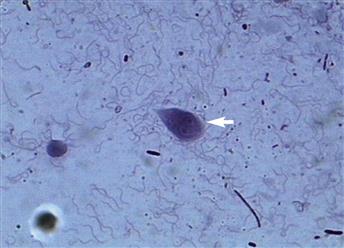
The motile trophozoite stage is pear-shaped and dorsoventrally flattened and possesses four pairs of flagella. This stage is 9 to 21 µm × 5 to 15 µm. Two nuclei and a prominent adhesive disc are present on the anterior portion of the trophozoite, suggesting to the observer that a pair of eyes is “staring back.” Figure 11-1 presents the motile trophozoite form of Giardia species.
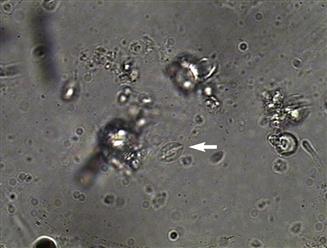
The mature cysts of Giardia species are oval and are 8 to 10 µm × 7 to 10 µm. They have a refractile wall and four nuclei. Immature cysts, which represent recently encysted motile forms, contain only two nuclei. Figure 11-2 presents a cyst of Giardia species. In dogs, diarrhea may begin as early as 5 days after exposure to Giardia, with cysts first appearing in the feces at 1 week.
 TECHNICIAN’S NOTE
The trophozoites of Giardia species are typically seen in animals with diarrhea rather than formed stool.
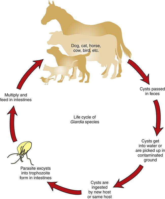
Recent advances in molecular biology have changed the way Giardia species are named. Currently, Giardia is referred to in terms of assemblages rather than species. Assemblages A and B are associated with human beings while assemblages C and D constitute the organisms found in dogs. Those organisms associated with cows, sheep, goats, horses, pigs, etc., are grouped in assemblage E. Assemblage F encompasses the forms found in cats while assemblage G includes the organisms found in rodents. While Giardia seems to be more species specific than once thought, assemblage A (that affecting humans) has been found in dogs and cats. This means there can still be a source of zoonotic infection from animals. However, it is more common that people are infected with Giardia by other people. Figure 11-3 shows the life cycle of Giardia species (Bowman, 2009).
Diagnosis of Giardia species is by standard fecal flotation. Zinc sulfate (specific gravity, 1.18) and Sheather’s sugar solution are considered the best flotation media for recovering cysts. Cysts are often distorted, with a semilunar appearance. The motile trophozoite occasionally can be found on direct smear of fresh feces using isotonic saline. Lugol’s iodine stain may be used to visualize the internal structures of both cysts and trophozoites. Fecal immunodiagnostic tests are also frequently used. Using immunodiagnostic tests in conjunction with fecal flotation tests will provide the best diagnostic protocol for diagnosing Giardia infection.
 TECHNICIAN’S NOTE
The cysts are hard to find in diarrheic samples, thus immunodiagnostic tests may be helpful with a definitive diagnosis.
Amoebae
Parasite: Entamoeba histolytica
Host: Canines, felines, primates, and humans
Location: Large intestines
Distribution: Worldwide but rare in the United States
Derivation of Genus: Internal amoeba
Transmission Route: Ingestion of cysts
Common name: Entamoeba
Entamoeba histolytica, the etiologic agent that produces amebic dysentery (an extremely severe dysentery) in humans may also produce sporadic infections in dogs. These cases usually have been acquired by association with infected humans. Although E. histolytica may produce acute or chronic diarrhea in dogs, it usually produces no pathology. Cats are rarely infected; most infections have been experimental.
E. histolytica occurs in two morphologic forms: a motile feeding stage (the trophozoite) and an environmentally resistant cyst stage. The motile trophozoite of E. histolytica ranges in size from 10 to 60 µm and has a single spherical nucleus that is 4 to 7 µm with a tiny pinpoint center, a structure called an endosome (Figure 11-4). The mature cysts are round and are 5 to 20 µm. They have a thin refractile wall and may demonstrate as many as four nuclei, each with its own endosome. Immature cysts, which represent recently encysted motile forms, contain only one nucleus (Figure 11-5).
 TECHNICIAN’S NOTE
Entamoeba histolytica is the parasite that causes amebic dysentery in humans.
Diagnosis of E. histolytica is by standard fecal flotation, which may be used to demonstrate both cyst and trophozoite forms. In a formed stool, usually the cyst form will be found; however, in diarrheic stool, both the cyst and the trophozoite forms may be observed. The direct smear technique may be used to observe motile forms in warm, freshly passed diarrheic stools. Lugol’s iodine stain may be used to visualize the internal structures of both cysts and trophozoites. Zinc sulfate may be used to concentrate cysts. If infection with E. histolytica is suspected in a human, a human pathology laboratory should be consulted for assistance with diagnosis. Because E. histolytica is primarily a human pathogen, great care should be taken with suspect feces.
E. histolytica also can be a significant problem in primates, such as monkeys and chimpanzees. Great care should be taken when handling suspect feces. Infection in monkeys has great public health significance.
 TECHNICIAN’S NOTE
E. histolytica is primarily a human parasite.
Ciliates
Parasite: Balantidium coli
Host: Canines and swine
Location: Cecum and colon of canines and large intestines of swine
Distribution: Worldwide
Derivation of Genus: Small bag
Transmission Route: Ingestion of cysts
Common Name: Balantidium
Balantidium coli is the ciliated protozoan found occasionally in the cecum and colon of dogs. This parasite has been associated with diarrhea in dogs. B. coli is more intimately associated with pigs (see the section on swine protozoan parasites).
Apicomplexans
Parasite: Cystoisospora species (formerly Isospora species)
Host: Canines, felines, and swine
Location: Small intestines
Distribution: Worldwide
Derivation of Genus: Equal seeds; sac same spore
Transmission Route: Ingestion of oocysts
Common Name: Coccidia
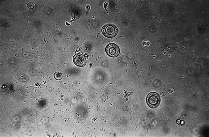
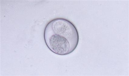
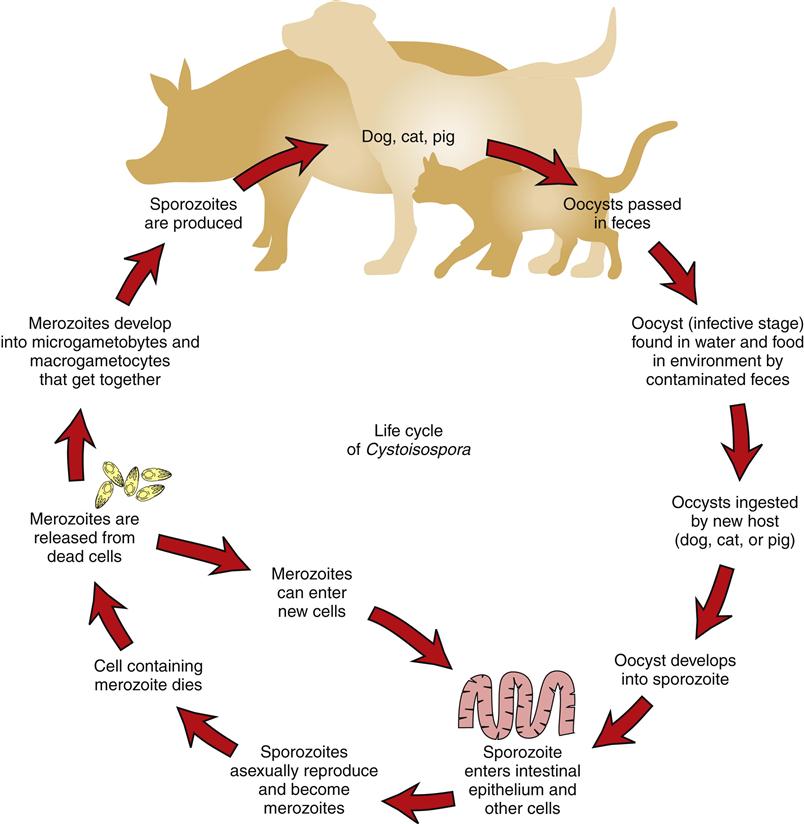
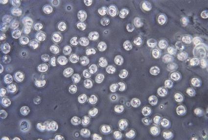
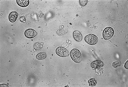
Cystoisospora species (coccidians) are protozoan parasites of the small intestine of dogs and cats. They produce a clinical syndrome known as coccidiosis, caused by inflammation of the intestine, one of the most commonly diagnosed protozoan diseases in puppies and kittens. It produces loose stool to a watery diarrhea. Coccidiosis is rarely a problem in mature animals. The oocyst is the diagnostic stage observed in a fecal flotation of fresh feces; it is unsporulated in fresh feces and varies in size and shape among the common Cystoisospora species. Figure 11-6 shows an unsporulated oocyst, and Figure 11-7 shows a sporulated oocyst. The canine coccidians and their measurements are Cystoisospora canis, 34 to 40 µm × 28 to 32 µm; C. ohioensis, 20 to 27 µm × 15 to 24 µm; and C. burrowsi, 10 to 14 µm × 7.5 to 9 µm. The feline coccidians and their measurements are Cystoisospora felis, 38 to 51 µm × 27 to 29 µm, and C. rivolta, 21 to 28 µm × 18 to 23 µm. Figure 11-8 shows the life cycle of Cystoisospora species. The prepatent period varies among species, but it is usually 7 to 14 days.
 TECHNICIAN’S NOTE
Cystoisospora spp. are some of the most commonly diagnosed of the most commonly diagnosed parasites in puppies and kittens.
Parasite: Toxoplasma gondii
Host: Definitive host is the feline. Can also occur in other animals and humans as incidental parasite.
Location: Intestines
Distribution: Worldwide
Derivation of Genus: Bow body
Transmission Route: Ingestion of sporulated oocysts
Common Name: Toxoplasma
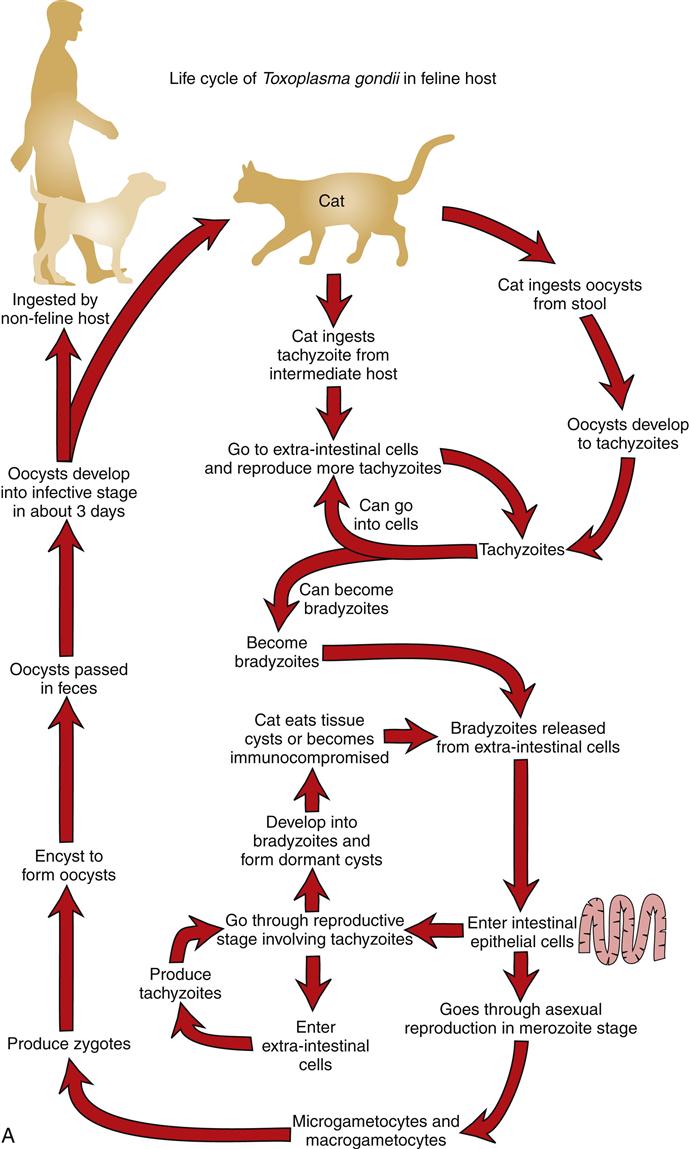
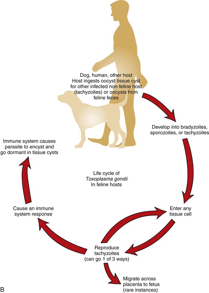
Toxoplasma gondii is an intestinal coccidian of cats. Its oocysts are usually diagnosed using a standard fecal flotation solution. Oocysts of T. gondii are unsporulated in fresh feces and measure 10 × 12 µm (Figure 11-9). Several immunodiagnostic tests using whole blood or serum are available for diagnosis of T. gondii. The prepatent period is highly variable, ranging from 5 to 24 days, and depends on the infection route. Figure 11-10A shows the life cycle of Toxoplasma gondii in the feline host and Figure 11-10B shows the life cycle in the nonfeline host. Because this parasite can use humans (and many other warm-blooded vertebrates) as intermediate hosts, T. gondii is a very important zoonotic parasite.
T. gondii can be a significant pathogen in humans (particularly the pregnant woman and her developing fetus). It is important to discuss this parasite with clients. Great care should be taken when handling suspect feces. Infection in cats has great public health significance.
 TECHNICIAN’S NOTE
The feline only sheds Toxoplasma gondii oocysts for up to 2 weeks of its life.
Parasite: Cyptosporidium species
Host: Canines, felines, ovines, swine, avians, guinea pigs, snakes, and mice
Location: Small intestine
Distribution: Worldwide
Derivation of Genus: Hidden small seeds
Transmission Route: Ingestion of oocysts
Common Name: Cryptosporidium, “Crypto”
Cryptosporidium species is another coccidian-like parasite that parasitizes the mucosal cells of the small intestine of a variety of animals and causes inflammation of those cells. These animals include cows, pigs, dogs, cats, birds, and guinea pigs. Cryptosporidium species in snakes, mice, and some species in cows parasitize the stomach and abomasum (cows). There is also a species of Cryptosporidium that parasitizes humans. In most cases of infection, diarrhea is the major clinical sign. The sporulated oocysts in the feces are oval to spherical and measure only 4 to 6 µm. Diagnosis is by standard fecal flotation. The oocysts are extremely small and may be observed just under the coverslip, not in the same plane of focus as other oocysts and parasite eggs (Figure 11-11). Examination of fresh fecal smears using special stains (modified acid-fast stain) is also helpful. ELISA tests are also being used to diagnose this parasite.
Because humans may become infected with Cryptosporidium species, feces suspected of harboring this protozoan should be handled with great care. Cryptosporidium is a zoonotic parasite. Cryptosporidium parvum is the most common zoonotic species. However, C. canis, C. felis, C. meleagridis (birds), C. muris (mice), and C. suis (pigs) have been seen in humans.
 TECHNICIAN’S NOTE
Feces suspected of containing Cryptosporidium oocysts should be handled with great care.
Parasite: Sarcocystis species
Host: Canines and felines
Location: Small intestine
Distribution: Worldwide
Derivation of Genus: Flesh cyst
Transmission Route: Ingestion of muscle of horses, pigs, and ruminants
Common Name: Sarcocystis
Sarcocystis species are the final coccidian parasites cited here that are found in the small intestine. Several species infect dogs and cats, and identification of an individual species can be quite difficult. The oocysts of Sarcocystis species are sporulated when passed in the feces. Each oocyst contains two sporocysts, each with four sporozoites. These individual oocysts measure 12 to 15 µm × 8 to 12 µm and may be recovered in a standard fecal flotation of fresh feces. The oocysts are ingested by a ruminant, horse, or pig depending on the specific species of Sarcocystis. The oocysts will excyst and go through multiple asexual reproductive stages before encysting in the muscles of the intermediate host. The muscle tissue of the intermediate host must be ingested to complete the maturation to the oocyte stage in the final host species, canine or feline.
 TECHNICIAN’S NOTE
There are seven species of Sarcocystis that infect dogs and five species that infect cats. Each of the species has a different intermediate host.
Circulatory System and Blood
Protozoans that infect the circulatory system and blood of dogs and cats are flagellates and apicomplexans.
Flagellates
Parasite: Trypanosoma cruzi
Host: Humans and occasionally canines
Location: Peripheral blood
Distribution: Primarily in Central and South America but occasionally in the southern United States
Derivation of Genus: Borer body
Transmission Route: Ingestion of intermediate host, reduviid bug, or feces of reduviid bug left on mucous membranes of final host
Common Name: Trypanosome
Trypanosoma cruzi is a hemoprotozoan (a protozoan found circulating in the peripheral blood). It is primarily found in Central and South America; however, it is occasionally reported in dogs in the southern half of the United States. This trypanosome is extracellular; it is not found within the red blood cell (RBC) itself. It “swims” within the blood. This swimming stage is called a trypomastigote and is 16 to 20 µm in length, approximately 3 to 10 times as long as an RBC is wide. It is banana-shaped and possesses a lateral, undulating membrane and a thin, whiplike tail (the flagellum) that is used for swimming. These parasites are also transmitted by blood-feeding arthropods called reduviid bugs, or “kissing bugs” (see Chapter 13). T. cruzi also has a resting cyst stage (the amastigote stage) that may be found encysted within cardiac muscle and other tissues such as the esophagus. The encysted amastigote stage lacks the undulating flagellum of the swimming trypomastigote stage.
The trypomastigote stage of T. cruzi may be found swimming within the peripheral blood and may be demonstrated in direct blood smears. The amastigote stage must be confirmed using histopathologic sectioning. Suspect tissues should be submitted to a histopathology laboratory for diagnosis by a veterinary pathologist or parasitologist.
 TECHNICIAN’S NOTE
The trypomastigote stage of Trypanosoma cruzi can be found in a peripheral blood smear.
Parasite: Leishmania species
Host: Canines
Location: Reticuloendothelial cells of capillaries and spleen as well as other internal organs and white blood cells
Distribution: Worldwide but rarely seen in North America
Derivation of Genus: Named after a famous physician, William Leishman
Transmission Route: Bite by an infective intermediate host, phlebotomine sand flies
Common Name: Leishmania
Leishmania species is another hemoprotozoan (blood protozoan). The parasite is primarily found in areas other than North America; however, it is occasionally reported in dogs that have traveled or been born overseas. It is rarely diagnosed in dogs that have never left the United States. This flagellate is intracellular and found within reticuloendothelial cells of the capillaries, spleen, and other internal organs and in monocytes, polymorphonuclear leukocytes, and macrophages. Instead of having a flagellum and being called the trypomastigote stage, as with T. cruzi, this parasite lacks the flagellum and is called the amastigote stage. Leishmania is transmitted by blood-feeding arthropods called phlebotomine sand flies (see Chapter 13).
The amastigote stage of Leishmania must be confirmed using histopathologic sectioning of infected organs. Suspect tissues should be submitted to a histopathology laboratory for diagnosis by a veterinary pathologist or parasitologist.
 TECHNICIAN’S NOTE
Leishmania species are rarely seen in dogs in North America.
Apicomplexans
Parasite: Babesia canis
Host: Canine
Location: Within red blood cells
Distribution: Europe, Africa, Asia, North and South America
Derivation of Genus: Named after a famous bacteriologist, Victor Babès
Transmission Route: Bite of an infective intermediate host, ticks
Common Name: Babesia or canine piroplasm
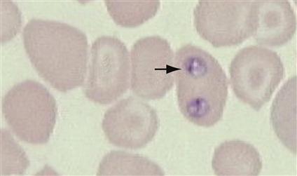
Babesia canis is an intracellular parasite found within the RBCs of dogs. It has been referred to as the canine piroplasm. The parasite demonstrates pear-shaped organisms within canine RBCs. This protozoan parasite is spread by the bite of infected ticks. Diagnosis is by observing basophilic, pear-shaped organisms within RBCs in stained blood smears (Figure 11-12). This parasite causes damage by residing within the red blood cells. Common symptoms include pale mucus membranes, icterus, hemoglobinuria, depression, hemoglobinemia, weakness, splenomegaly, fever, and anorexia.
 TECHNICIAN’S NOTE
Babesia canis can be broken down into three subspecies that denote in which country the species is found. B. canis canis is found in Europe, B. canis vogeli is found in Northern Africa and North America, and B. canis rossi is found in Southern Africa (Bowman, 2009).
Parasite: Cytauxzoon felis
Host: Feline
Location: Within red blood cells
Distribution: Africa and United States
Derivation of Genus: Hollow vessel-increasing animal
Transmission Route: Bite from an infective intermediate host, tick
Common Name: Cytauxzoon
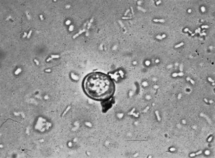
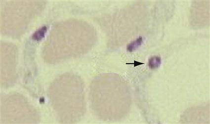
Cytauxzoon felis is another intracellular parasite that has been reported in the RBCs of cats in sporadic sites (Missouri, Arkansas, Georgia, and Texas) throughout the United States and Africa. It also produces piroplasms; however, these bodies have been described as being in the shape of a “bejeweled ring” and are referred to as the ring form in stained blood smears (Figure 11-13). This disease is spread by ticks, and its prognosis is poor. Diagnosis is made by observing the bejeweled-ring piroplasms within the RBCs in stained blood smears. Common symptoms include fever, icterus, anemia, dehydration, and death. As with Babesia canis, Cytauxzoon felis causes damage by inflammation within the red blood cells.
 TECHNICIAN’S NOTE
Cytauxzoon felis is diagnosed by finding the bejeweled ring form in the RBCs in stained blood smears.
Parasite: Hepatozoon canis and Hepatozoon americanum
Host: Canines
Location: Gamonts are within white blood cells while schizonts are found in the endothelial cells of the spleen, bone marrow, and liver.
Distribution: United States, Africa, Asia, and Southern Europe
Derivation of Genus: Liver animal
Transmission Route: Ingestion of infective intermediate host, Rhipicephalus sanguineus tick for H. canis and Amblyomma maculatum tick for H. americanum
Common Name: Hepatozoon
Hepatozoon canis and Hepatozoon americanum are intracellular, malaria-like parasites affecting dogs. The blood forms (the gamonts) of these protozoan parasites are found in the leukocytes. (Leukocytes containing gamonts of H. canis are common in peripheral blood smears, whereas those of H. americanum are rare.) Schizonts are found in the endothelial cells of the spleen, bone marrow, and liver. The gamonts are surrounded by a delicate capsule and stain pale blue with a dark, reddish purple nucleus. Numerous pink granules are found in the cytoplasm of the leukocyte. The “onion skin” tissue cysts of H. americanum are found in skeletal muscle of dogs (Figure 11-14). This is an unusual parasite in that the dog becomes infected by ingestion of an infected tick, Amblyomma americanum. H. canis is well adapted to its canine host and varies from being subclinical to producing mild disease. H. americanum produces a violent and frequently fatal course of disease; it is theorized to have crossed the species barrier from a wild animal host to the domestic dog.
 TECHNICIAN’S NOTE
Almost all intracellular malaria-like parasites are transmitted by the bite of an arthropod (a mosquito or a mite), but Hepatozoon species is spread by the ingestion of an arthropod.
Ruminants (Cattle and Sheep)
Gastrointestinal Tract
Protozoans that infect the gastrointestinal tract of ruminants are ciliates and apicomplexans.
Ciliates
Nonpathogenic ciliates live in the rumen of all ruminants, including cattle and sheep. These are mutualists; in this type of symbiotic relationship, both individuals benefit from the association. The cow provides a warm, moist, particulate medium in which the ciliates live; the ciliates, in exchange, aid in digestion by breaking down cellulose. Sometimes, ciliates may be observed in the diarrheic feces of cattle and sheep. These ciliates may be recovered on fecal flotation. Similar ciliates occur in the cecum of the horse and are often recovered from diarrheic feces.
Apicomplexans
Parasite: Eimeria species, Eimeria bovis, and Eimeria zuernii
Host: Ruminants
Location: Cecum and colon
Distribution: Worldwide
Derivation of Genus: Named after a famous zoologist, Gustav Eimer
Transmission Route: Ingestion of oocysts
Common Name: Coccidia
Ruminants serve as host to many species of the coccidian parasite Eimeria. It is often difficult to identify the individual species of Eimeria because their oocysts are similar in size and shape. The life cycle is similar to that of Cystoisospora species (see Figure 11-8) and causes damage by inflammation of the intestinal cells. The two most common species of coccidia in cattle are Eimeria bovis and Eimeria zuernii; they can be differentiated on a standard fecal flotation. Oocysts of E. bovis are oval, have a micropyle (opening), and measure 20 × 28 µm; the oocysts of E. zuernii are spherical, lack the micropyle, and measure 15 to 22 µm × 13 to 18 µm. When oocysts are recovered on fecal flotation, the observation is usually noted as coccidia. The coccidian oocysts of ruminants can be partially differentiated by size and appearance and cause diarrhea and possibly neurologic signs, such as muscle tremors, convulsions, and blindness (Figure 11-15).
 TECHNICIAN’S NOTE
Individual species of Eimeria can be partially differentiated by oocyst size and appearance.
Parasite: Cryptosporidium species (discussed in greater detail in canine and feline section)
Host: Canines, felines, bovines, swine, avians, guinea pigs, snakes, and mice
Location: Small intestine
Distribution: Worldwide
Derivation of Genus: Hidden small seeds
Transmission Route: Ingestion of sporulated oocysts
Common Name: Cryptosporidium; “crypto”
Cryptosporidium species is another coccidian that parasitizes the small intestine of a variety of animals, including cattle, sheep, and goats. The sporulated oocysts in the feces are colorless and transparent and measure only 3 to 5 µm. Diagnosis is by standard fecal flotation and stained fecal smears. Because humans may become infected with Cryptosporidium species, feces suspected of harboring this protozoan should be handled with great care. These oocysts are often recovered using Sheather’s sugar solution. (See Figure 11-11 for features of the oocysts of Cryptosporidium species.)
 TECHNICIAN’S NOTE
Feces suspected of being infected with Cryptosporidium should be handled with great care due to the potential for zoonosis.
Stay updated, free articles. Join our Telegram channel

Full access? Get Clinical Tree


