CHAPTER 64 Clinical Reproductive Anatomy and Physiology of the Buck
ANATOMY OF THE REPRODUCTIVE TRACT
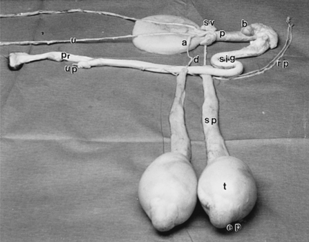
The male reproductive organs include a pair of testes, which produce sperm and hormones; a pair of excurrent ducts (rete testis, ductuli efferentes, epididymis, and ductus deferens) where sperm mature and then are stored (in the tail of the epididymis) until ejaculated; a penis, which serves as a copulatory organ; and a set of accessory sex glands whose secretions (seminal plasma) form the bulk of the semen (Fig. 64-1).
The testes, although developed in the abdomen, migrate prenatally through the inguinal canal into the scrotum. The mature testes are ovoid in shape and positioned vertically in the scrotum; each testis can weigh 90 to 100 g in a 2- to 3-year-old Nubian buck with a 28 to 30 cm scrotal circumference.1 The rete testis, a network of intercommunicating channels, occupies almost two thirds of the central axis of the testis. The ductuli efferentes, a group of 15 to 20 tubules, connect the rete testis with the epididymis, which, although a single tube about 60 to 80 m long, can be divided into three main regions: head, body, and tail. The head lies on the dorsocaudal border of the testis, the body is a thin strip-like structure attached on the caudomedial border of the testis, and the tail is an enlarged promontory structure located on the ventromedial border of the testis that can be palpated through the scrotum in mature goats. The ductus deferens connects the tail of the epididymis to the pelvic part of the urethra. Enwrapped by its own peritoneal fold, the ductus deferens ascends through the inguinal canal to enter the abdominal cavity at the vaginal ring. It then separates from the other parts of the spermatic cord and pursues a course via the genital fold to the caudal part of the bladder. The distal part of the ductus deferens enlarges to form the ampulla, which is 6 to 7 cm long and 4 to 5 mm in diameter, and dips under the prostate to open with the excretory duct of the seminal vesicle on the side of the colliculus seminalis.2
Seminal vesicles, the largest of the accessory sex glands, are elongated saccular organs with a lobulated surface. They lie on each side of the caudal part of the dorsal surface of the bladder. Each gland in a 2- to 3- year-old Nubian buck is about 4 cm long, 2.5 cm wide, and 1.5 cm thick.2 Although they lie in close contact with the rectum, it is difficult to palpate them through the rectal wall. Each excretory duct passes through the prostate gland and opens on the colliculus seminalis. The entire prostate gland is disseminated and completely surrounds the wall of the pelvic urethra. Numerous small ducts carry the prostatic secretion into the urethra. The paired bulbourethral glands lie on the dorsal surface of the urethra opposite the ischial arch and are covered by a thick layer of dense connective tissue and by the proximal part of the thick bulbospongiosus muscle. The excretory duct of each gland opens into the urethra under a blind fold of mucous membrane, which makes it difficult to pass a catheter into the bladder.
SPERMATOGENESIS AND TRANSPORT, MATURATION, AND STORAGE OF SPERM
Spermatogenesis
Spermatogenesis is a highly synchronized and hormonally controlled sequence of events wherein the germ cells undergo a series of divisions and differentiation (spermatogonia, primary spermatocytes, secondary spermatocytes, early spermatids, and late spermatids) resulting in the formation of haploid sperm. Mathematically, 64 sperm cells can result from one spermatogonium, and in sheep, the entire cycle of spermatogenesis, from the first spermatogonial division to the release of sperm into the lumen of the seminiferous tubule, is completed in 49 days.3 The cycle is expected to be similar in goats. Spermatogenic cycling begins with the onset of puberty and is repeated at a fixed interval throughout the life of the animal.
The differentiation of spermatogonia to sperm is closely regulated by Sertoli cells, which form close junctional contacts with germ cells. In addition to Sertoli cell secretions, testosterone is essential for spermatogenesis in all mammalian species, including goats. In fact, spermatogenesis requires a 50 to 100 times higher level of testosterone surrounding the germ cells than in the systemic circulation.4 Testosterone is also essential for production of seminal plasma, epididymal maturation of sperm, and development and maintenance of secondary sex characteristics and libido. More than 90% of the body’s testosterone is secreted by the Leydig cells, whose structure and function depend on the functional maturity of the hypothalamus-pituitary–Leydig cell axis. The hypothalamus secretes luteinizing hormone–releasing factor, which stimulates the pituitary to secrete luteinizing hormone, which, in turn, stimulates the Leydig cells to secrete testosterone. The body maintains a homeostatic level of testosterone through the negative feedback effects of testosterone on the pituitary gland or the hypothalamus or both.4
Stay updated, free articles. Join our Telegram channel

Full access? Get Clinical Tree


