CHAPTER 30 Clinical Chemistry of the Puppy and Kitten
Neonates have decreased functional capacity of many organ systems because of incomplete development of these organs at birth. As they age, organ function increases and variations in enzyme levels and the products related to normal metabolism, filtration, and function of these organs will change in accordance to appropriate growth of the animal. Serum chemistry can be useful to detect often subtle abnormalities of these organ systems, and although reference ranges have been established for the normal parameters of most of these analytes, these values have often been obtained from adult animals. Because of variations in enzymology and functional capacity of neonatal organ systems, care must be taken when interpreting any changes in chemistry values when using standard adult reference ranges.
This chapter focuses on the typical development and acquisition of normal biochemical constituents in puppies and kittens and illustrates the many differences between adult and neonatal biochemical parameters. Although published reference ranges are provided (Tables 30-1 to 30-5) based on the available research in current literature, it is recommended that reference intervals be established for each laboratory because of the lack of standardization among reference laboratories. Practitioners, however, may use these ranges as guidelines for interpretation of serum biochemical results in puppies and kittens aged less than 1 year. Hereditary conditions affecting biochemical parameters of young dogs and cats are shown in Table 30-6.
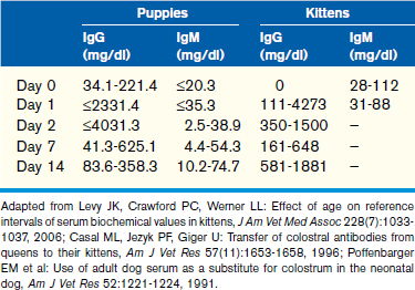
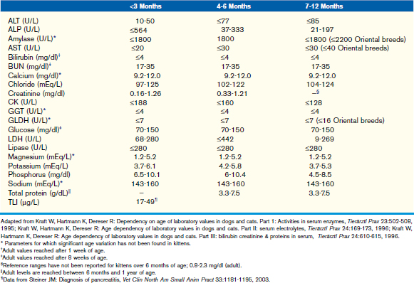
TABLE 30-6 Hereditary conditions affecting biochemical parameters of young dogs and cats
Condition | Mode of Inheritance |
---|---|
Benign familial hyperphosphatasemia in Siberian Huskies | Likely autosomal (exact mode of inheritance not known) |
Musculodystrophy | X-linked |
Hyperchylomicronemia in cats | Believed to be autosomal recessive |
Pancreatic acinar atrophy and exocrine pancreatic insufficiency in German Shepherd Dogs | Autosomal recessive |
Severe combined immunodeficiency | |
Basset Hounds | X-linked |
Jack Russell Terriers and Cardigan Welsh Corgis | Autosomal recessive |
IgA deficiency | Unknown |
Hypercholesterolemia in Rottweilers and Dobermans | Unknown |
Hereditary renal dysplasia in Lhasa Apsos | Autosomal recessive |
Congenital dwarfism | Autosomal recessive |
Hyperkalemia in posthemolysis in Akitas, Japanese Shibas, and Jindos | Believed to be autosomal recessive |
Glucose
Glucose in the blood is closely regulated and normally maintained by three major mechanisms: intestinal absorption, hepatic production, and, to a lesser degree, renal production. In young animals with reduced development of normal organ, mainly hepatic, function, there is reduced potential for gluconeogenesis and glycogenolysis. Therefore, much of the plasma glucose concentration in young animals is obtained via ingestion, which makes neonates particularly sensitive to hypoglycemia during incidents of stress, illness, and reduced intake.
In puppies, glucose levels are lowest immediately after birth and then significantly increase after approximately 3 days with normal suckling. No significant variations are noted in glucose from day 8 until post-nursing. Glucose concentration then gradually decreases over time and levels off to normal adult levels at approximately age 9 months. The lower glucose values identified in young puppies, immediately after birth, are likely caused by insufficient blood sugar regulation feedback mechanisms and decreased hepatic functional capability.
In contrast to puppies, glucose levels have been shown to be higher immediately after birth in kittens with a gradual decline throughout the growth period to reach normal adult concentrations soon after weaning.
Hypoglycemia
Decreased glucose absorption secondary to starvation, maldigestion, and poor nursing or agalactia of the queen or bitch are the most common causes of hypoglycemia in neonates. Disease states such as diarrhea, dehydration, or hypothermia may exacerbate hypoglycemia in these patients. A syndrome known as transient juvenile hypoglycemia is well recognized and of particular concern in miniature and toy breeds of dogs; therefore, special consideration must be taken to inhibit decreased intake in these breeds postweaning. Inadequate glycogen and protein stores, decreased gluconeogenesis resulting from decreased hepatic function, and suboptimal epinephrine-mediated response to hypothermia and hypoglycemia are implicated in this syndrome.
Sepsis is another significant cause of hypoglycemia in animals. Neonates may be more susceptible to sepsis as a result of inadequate developing immune function and the inability to rely on normal mobilization of glycogen and protein stores as previously mentioned.
Liver dysfunction should always be considered in cases of persistent hypoglycemia in puppies and kittens. This may include primary causes such as portosystemic shunts (PSSs) in either species or acquired dysfunction secondary to infectious disease, for example, hepatitis in puppies or feline infectious peritonitis (FIP) in kittens.
Common causes of hypoglycemia in neonates are listed in Box 30-1.
Hyperglycemia
Hyperglycemia secondary to excitation, stress, or fear resulting from transient catecholamine release is common in cats and dogs regardless of age but may be particularly pronounced in cats. Likewise, chronic stress resulting in cortisol-induced gluconeogenesis is also relatively common in small animals and may accompany a variety of disease states. Postprandial hyperglycemia may occur in puppies and kittens within 1 to 4 hours after digestion of a meal.
Certain drugs may cause transient hyperglycemia in animals, including neonates, after administration. These compounds include anesthetizing agents such as medetomidine, xylazine, and ketamine, as well as analgesics such as morphine, fentanyl, and butorphanol, commonly used for elective procedures such as spays and neuters of puppies and kittens.
Juvenile or type 1 diabetes mellitus is more common in young dogs, and it has not been well documented in feline medicine. Juvenile diabetes is discussed in more detail elsewhere in this text but should be suspected in puppies with persistent hyperglycemia, glucosuria, and clinical signs of disease (see Chapter 45).
Common causes of hyperglycemia in neonates are listed in Box 30-1.
Protein
Serum proteins can be divided into two major categories: albumin and globulins. Albumin and the majority of globulins are synthesized by the liver in response to cytokine stimulation. Nutritional intake of proteins may also affect serum concentrations. Plasma also contains coagulation proteins that are also, primarily, produced by the liver. The majority of these coagulation proteins are not present in serum samples as they have been “consumed” by clot formation.
Age-associated increase of total protein and albumin is well described in animals. These increases are attributed to normal immune stimulation resulting in an elevated globulin fraction and increased albumin production resulting from improved liver function and intestinal absorption. As a result, in mammals, total protein concentrations are low at birth, increase dramatically after absorption of colostrum, and then decrease over 1 to 5 weeks as colostrum is metabolized. Total protein concentration then gradually increases to achieve adult levels within 6 months to 1 year.
Albumin
Albumin has a half-life of approximately 8 days in dogs and cats. It accounts for approximately 75% to 80% of the colloidal osmotic activity of plasma and is also a negative acute phase protein. It is important in the transport of many endogenous and exogenous molecules, including hormones, unconjugated bilirubin, and several drugs.
Globulins
Globulins are further classified into alpha (α)-, beta (β)-, and gamma (γ)-fractions. Most acute phase proteins are either α- or β-globulins, and, depending on the inflammatory process and type of globulin, variable increase and decrease of certain globulins may aid diagnosis of disease, response to therapy, and prognosis. The γ-globulin fraction contains the immunoglobulins of which IgG, IgM, IgE, and IgA are measurable in serum.
Puppies are born hypogammaglobulinemic with only a small amount of IgG and IgM and no detectable IgA in serum at birth. Therefore, total protein concentration in puppies is initially low, particularly precolostral intake. Protein concentration then steadily increases during the first year of life and is stable from age 1 year onward. Decreased total protein in puppies aged 8 weeks or less is likely caused by inefficient ability of the liver to synthesize it. Therefore, there is no compensation for increased blood volume that occurs during nursing and inadequate reabsorption ability from metabolites in the gastrointestinal tract. Puppies may also have higher intravascular water content than adults, resulting in a dilutional effect of their serum analytes.
In puppies, 5% to 10% of maternally derived IgG, and possibly IgM, may be transferred transplacentally. The remainder of IgG is transferred in the colostrum, resulting in an initial spike in globulins immediately after colostral ingestion. Because IgM circulates as a pentamer, it is unlikely that IgM is absorbed through the colostrum because of the large size of the molecule. This is supported by studies showing that puppies given adult canine serum subcutaneously at birth have higher IgM concentrations than puppies given the same serum orally. Additional information pertaining to passive transfer of maternal antibodies can be found in Chapter 2. The half-life of IgG and IgA in puppies is 10 days and 4 to 5 days, respectively. Therefore, a decrease in globulins and an increase in albumin within the first 6 weeks of life are identified as a result of degradation of maternally derived antibodies and increased synthesis of albumin as normal liver function develops. This leads to a peak in the albumin to globulin (A/G) ratio at 6 to 8 weeks, after which the A/G ratio decreases, consistent with a slight increase in globulin fraction likely related to maturation of the immune system.
In kittens, approximately 25% of serum immunoglobulin concentration in newborns is attributable to transplacental absorption. However, unlike in puppies, IgG and IgA are not present in kitten serum at birth. Colostral absorption of maternal antibodies occurs within a very short timeframe in kittens. It is detected at 12 hours after birth but does not occur at 16 hours after birth. After colostral absorption, IgG and IgA concentrations peak within the first day of life and then decrease steadily as maternal antibodies decrease. The half-life of maternally derived IgG and IgA in kittens is 4 days and 1 to 2 days, respectively. Postweaning, IgG increases gradually as the kitten’s production capacity by the developing immune system increases. IgA increases at a slower rate than IgG but then increases markedly at age 6 weeks. Unlike IgG and IgA, IgM may be detected in small amounts at birth (28 to 112 mg/dl), and, similar to other immunoglobulins, it increases steadily after the first few days of life.
Hypoproteinemia
Panhypoproteinemia may occur with gastrointestinal disorders, blood loss, liver dysfunction, or renal disease. However, it is important to note that many of these diseases may result from infectious or inflammatory causes resulting in increased globulins secondary to inflammation, thus masking an underlying loss of protein.
Disorders involving the intake and assimilation of protein are common causes for hypoproteinemia in animals. Intestinal malabsorption may result in a protein-losing enteropathy as both albumin and globulins leak through the intestinal wall into the lumen and are then digested and excreted. This may result from a variety of inflammatory conditions and is commonly associated with intestinal parasites in young animals, particularly those causing blood loss (e.g., whipworms). Exocrine pancreatic insufficiency results in the maldigestion of nutrients, including proteins, because of decreased production of digestive enzymes by the pancreas, in this case, trypsin. Because the liver uses amino acids obtained from protein digestion for the production of albumin, malnutrition or maldigestion of proteins results in decreased production of albumin despite normal liver function.
Severe liver dysfunction may result in decreased production of all proteins; however, hypoalbuminemia is typically detected primarily.
Protein-losing nephropathy may result in a net loss of both albumin and low molecular weight globulins because of defective protein tubular resorption of these molecules. Likewise, glomerular disease may result in a generalized loss of protein fractions.
Blood loss or hemorrhage secondary to trauma, coagulation disorders, including warfarin and brodifacoum intoxication, viral disease (e.g. parvovirus), and intestinal parasitism (mentioned previously) results in proportional loss of all blood constituents, including protein. In response to blood loss, interstitial fluid moves into the vascular space to increase blood volume. This results in dilution of the remaining blood constituents and further decreases total protein concentrations in both serum and plasma.
Hypoglobulinemia is most commonly associated with a failure of passive transfer in young animals, including puppies and kittens. As previously mentioned, there is a narrow window for absorption of immunoglobulins by the gut in animals (less than 24 hours). Failure of passive transfer is typically diagnosed by measuring IgG in serum via radial immunodiffusion (RID) method. An enzyme-linked immunosorbent assay is also available but is not as commonly used for measuring IgG in puppies and kittens because of difficulties in interpretation of results.
An inherited deficiency of B lymphocytes has been reported as an X-linked trait in Basset Hounds and also as an autosomal recessive trait in Cardigan Welsh Corgis and Jack Russell Terriers (see Table 30-6). This syndrome, known as severe combined immunodeficiency syndrome (SCID), results in a marked decrease in immunoglobulin production and occurs with profound lymphopenia in affected puppies. Selective immunoglobulin deficiencies have also been identified in other breeds of puppies, including IgA deficiency in German Shepherd Dogs, Beagles, and Shar-Peis (see Table 30-6).
Hyperproteinemia
Increased hepatic synthesis of albumin does not occur; therefore, hyperalbuminemia, often accompanied by hyperglobulinemia, is usually caused by hemoconcentration secondary to dehydration. Hyperglobulinemia is commonly seen with inflammation and is caused by both increased production of immunoglobulins and increased production of acute phase proteins. In dogs, C-reactive protein has been found to be the most common acute phase protein and is a sensitive, although nonspecific, indicator of inflammation in this species, often preceding changes in the leukocyte profile.
Kittens with FIP commonly exhibit hyperproteinemia secondary to hyperglobulinemia. In kittens with the wet form of FIP, an A/G ratio greater than 0.8 in effusions has been shown to be predictive for ruling out FIP. Similar to C-reactive protein in dogs, the acute phase protein α1-acid glycoprotein (AGP) is a very sensitive indicator of inflammation in cats. AGP levels greater than 1 to 5 g/L (RR < 0.48 g/L) in serum, plasma, or effusion samples are also found to aid in distinguishing cats with FIP from cats with other diseases with similar clinical signs.
Lipids
Lipids in plasma are divided into five categories: cholesterol, cholesterol esters, triglycerides, phospholipids, and long-chain fatty acids (LCFAs). LCFAs are obtained through the diet where they are absorbed from the digestive tract and then are incorporated into triglycerides by the intestinal epithelial cells. They may also be synthesized by the liver, adipose tissue, and mammary glandular tissue from glucose. Lipids are transported throughout the body attached to proteins. LCFAs combine with albumin for transport to tissues, whereas triglycerides and cholesterol attach to proteins, forming lipoproteins. These lipoproteins are broken down into very-low-density lipoproteins (VLDL), low-density lipoproteins (LDL), intermediate-density lipoproteins (IDL), and high-density lipoproteins (HDL), which can be differentiated by density and electrophoretically. The function of these lipoproteins is to transport aqueous insoluble lipids (e.g., cholesterol and triglycerides) through blood to tissues for metabolism, storage, or secretion. Cholesterol may be synthesized by the liver, or it may be absorbed from the intestine by animals eating animal protein. Cholesterol is predominantly transported by LDLs and HDLs in the bloodstream and is cleared from the serum by uptake by the liver. In the liver, cholesterol may be converted into bile acids or excreted directly into the bile.
Neonates are predominantly reliant on dietary absorption of lipids because of their decreased liver capacity to synthesize triglycerides and cholesterol. Nursing is an important source of lipids in neonates as milk contains a high content of fat, thus providing a high-calorie form of energy. In fact, plasma triglycerides have been shown to increase by twofold within 4 hours of a fat-rich meal. As a result, cholesterol is higher in nursing animals versus adults because of the high fatty acid content in milk. On ingestion of dietary fat, fatty acids are converted to triglycerides and transported through the blood in chylomicrons. Similarly, triglycerides formed by the liver are transported by VLDLs. Cellular uptake by myocytes and adipocytes for storage and transfer of lipids by lipoproteins is facilitated by interaction with several key enzymes and cell surface receptors that regulate the flux of lipids and the concentration of lipoproteins within plasma.
In puppies, cholesterol and triglyceride concentrations are highest at less than 8 weeks of age because of ingestion of milk fat during nursing. Postweaning, cholesterol and triglyceride concentrations gradually decrease but may peak again at age 5 to 6 months. After 6 months, values typically decrease until adult levels are reached.
For kittens, triglyceride and cholesterol concentrations are highest in nursing kittens, and upper reference limits in these kittens have been suggested to include triglyceride values up to 963 mg/dl and cholesterol values up to 521 mg/dl. These concentrations begin to decrease postweaning, because of withdrawal of milk consumption, to reach adult values by approximately age 9 to 12 months. Cats have been shown to have 5 to 6 times the levels of HDL versus LDL for lipid transport. Certain lipoproteins, particularly LDL, have been shown to decrease in preadolescent kittens (approximately 20 weeks of age) and are significantly lower than in adolescents (age 9 to 12 months). This is likely because of increased uptake of cholesterol to meet the needs of rapid tissue growth, sexual development, and steroidogenesis.
Hypolipidemia
Starvation or malnutrition, including poor nursing ability, in neonates is a serious cause of hypolipidemia, particularly hypotriglyceridemia. As the majority of lipids are obtained via ingestion of milk fats in these animals, lipid stores are rapidly depleted, and severe energy imbalances may ensue. Maldigestion and malabsorption of fats secondary to gastrointestinal disease may result in hypotriglyceridemia and, possibly, hypocholesterolemia in animals. Syndromes such as pancreatic insufficiency and inflammatory bowel disease with lymphangiectasia or the presence of gastrointestinal parasites may be responsible for marked decreases in serum and plasma lipid concentrations.
Hypocholesterolemia is commonly associated with liver dysfunction, as the liver is the major site of cholesterol synthesis. Disease may be acquired, secondary to infectious causes, toxic insult, or hypoxia, or it may be associated with a congenital defect such as PSS.
Hyperlipidemia
Hyperlipidemia occurs most commonly in young and adult animals postprandially as a result of increasing triglyceride concentrations after gastrointestinal absorption. To avoid this syndrome, a 12-hour fast before sampling is recommended in adults to minimize the amount of circulating chylomicrons, which may turn the serum milky white and lyse red blood cells, thus adversely affecting laboratory evaluation of both biochemical parameters and blood counts. However, this practice may be risky in young animals, particularly in miniature and toy breeds of puppies because of their propensity for developing hypoglycemia as mentioned previously.
Although liver failure is associated with decreased production of cholesterol in animals, other forms of liver disease may be accompanied by hyperlipidemia, particularly in disorders associated with cholestasis. Because the liver is the major route of cholesterol secretion, hypercholesterolemia and hypertriglyceridemia may result from decreased hepatic uptake and excretion of cholesterol into the bile and decreased lipoprotein production.
In animals with nephrotic syndrome, renal loss of albumin stimulates production of cholesterol and, occasionally, triglycerides in an attempt to increase plasma oncotic pressure.
Primary/congenital hyperlipidemia is described in dogs and cats (see Table 30-6). For example, primary hyperlipidemia secondary to hypercholesterolemia with normotriglyceridemia has been reported in some dog breeds such as Doberman Pinschers and Rottweilers. Fasting hypertriglyceridemia (>5 mmol/L or >454.54 mg/dl) with severe hemolytic anemia may be associated with idiopathic hyperlipidemia in kittens, a congenital condition in cats caused by a defect in lipoprotein lipase, the enzyme necessary for cellular uptake of triglycerides by myocytes and adipose. Elevated chylomicrons and VLDLs with or without low LDL and HDL are also identified in affected kittens. Clinical signs of this syndrome include weakness, inappetence, peripheral neuropathy (hindlimb paralysis), retinal disease (lipemia retinalis), tachycardia, and tachypnea resulting from anemia.
Congenital hypothyroidism with hypercholesterolemia has been reported in puppies and kittens. The mechanism is not completely understood; however, it may be associated with decreased hepatic metabolism of lipids and decreased fecal excretion of cholesterol. This disorder is discussed in more depth in Chapter 45. Some common causes for hyperlipidemia are summarized in Box 30-3.

Stay updated, free articles. Join our Telegram channel

Full access? Get Clinical Tree

