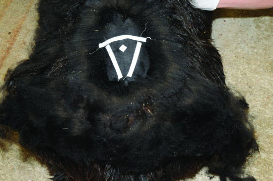
LS taps are safer than AO taps and may be performed in standing animals, animals in sternal recumbency, or animals in lateral recumbency. The flow of CSF courses from cranial to caudal, and LS taps are therefore useful for intracranial or spinal disease. With the animal standing or in the cushed position with the hind limbs flexed to open the space, the operator locates the wide LS space between the ileal wings (Figure 66.2). An easy method to select the site of penetration of the skin is to find the most caudal palpable dorsal spinous process of the lumbar vertebra and insert the needle approximately 3 to 4 cm caudal to this point and directly on dorsal midline.
Figure 66.2 Site for lumbosacral CSF tap. The animal is in sternal recumbency and restrained manually or with sedation. Cranial is to the top of the photo. The cranial piece of tape marks the level of the ileal wings. The target site for needle insertion is slightly caudal to the level of the ileal wings, where there is a palpable widening of the intervertebral space.
The area for CSF tap should be surgically clipped and prepared and 0.5 mL of 2% lidocaine injected subcutaneously to aid in the procedure. A 20- or 18-gauge × 3.5-inch (8.9-cm) needle with stylette is inserted in the LS space, perpendicular to the spine (Figure 66.3). If the back is arched, care must be taken to be perpendicular to the spine and not to the ground. The needle is advanced between the vertebrae until a slight pop is felt as the needle passes through the interarcuate ligament (dorsal spinous ligament). As the needle is advanced into the subarachnoid space, a second, more subtle “pop” may be felt. The second “pop” is often associated with a sudden reflex movement activity, and an assistant should provide effective restraint in anticipation of this reaction. At that time, the stylette is removed (Figure 66.4) and wiped across the operator’s sterile gloved hand. If moisture is present on the stylette, CSF may be aspirated (Figure 66.5).
Figure 66.3 Insertion of the spinal needle into the lumbosacral space. The needle is grasped with sterile gloves both at the hub and near the skin in order to stabilize and guide the needle.
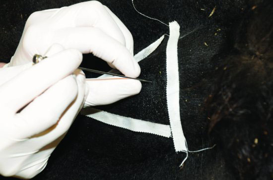
Figure 66.4 Once the needle is suspected to be in the subarachnoid space, the stylette is removed, the needle stabilized, and the hub of the needle is observed for flow of CSF. If none is seen, the stylette may be wiped across the back of the operator’s sterile glove to check for moisture, then aspiration is performed. If no fluid is obtained, the stylette is replaced and the needle repositioned.
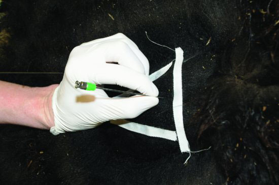
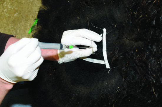
Figure 66.5 A sterile syringe is used to aspirate the CSF sample. Slow, steady aspiration should be performed with the needle stabilized to minimize trauma.
Stay updated, free articles. Join our Telegram channel

Full access? Get Clinical Tree


