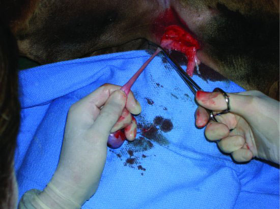
Figure 64.2 Initial midline incision into the prescrotal region. The operator’s left thumb is pushing one testicle up and maintaining its position under the incision to protect the penis from accidental incision.
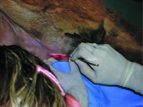
Figure 64.3 Pressure is applied under the testicle into the incision, and the incision is extended until the testicle (with tunic intact) is exteriorized.
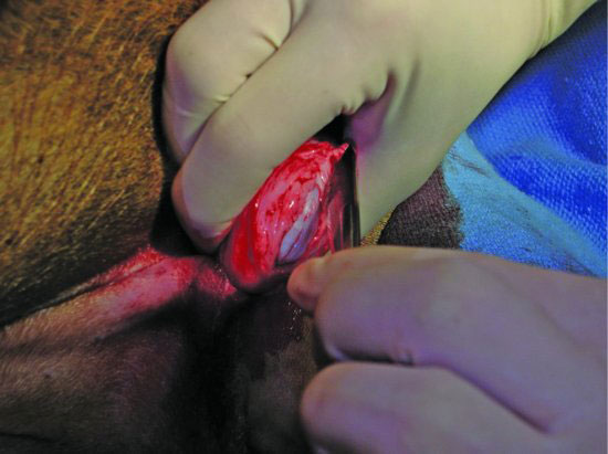
Figure 64.4 The testicle, tunic, and scrotal fat are exteriorized, and the spermatic cord is isolated.
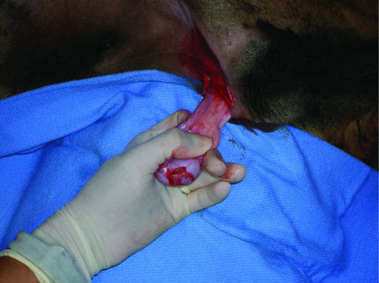
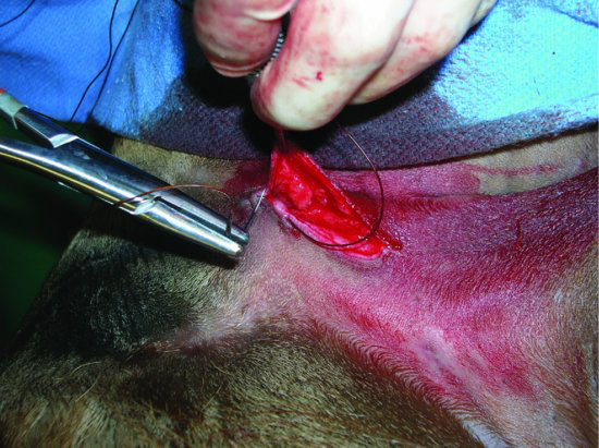
Figure 64.6 Absorbable suture material is used to place a transfixing or encircling ligature around the spermatic cord.
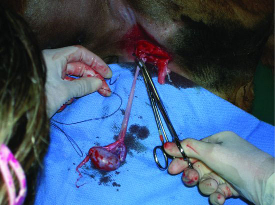
Figure 64.7 Both testicles have been removed, and the single prescrotal incision remains. Some operators will leave this incision open to heal.
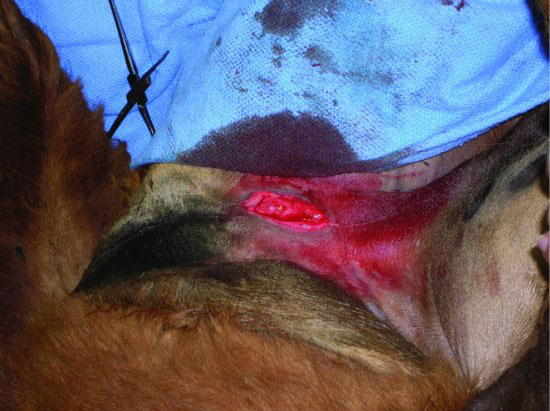
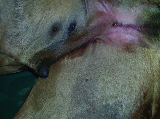
Figure 64.9 The completed, closed prescrotal castration results in an aesthetically pleasing surgical site that is not excessively attractive to flies.
Stay updated, free articles. Join our Telegram channel

Full access? Get Clinical Tree


