Case Descriptions

Chapter 21 Case Descriptions


This chapter was written for the second edition of this textbook, and despite the change for this third edition to a case-based format throughout the book, we decided to retain most of this chapter in its original form and have added a few new bonus cases. Consider this chapter a test of what you have learned from the preceding chapters. The format is the same as that used for the case examples throughout the book. You must appreciate that magnetic resonance (MR) and computed tomographic (CT) imaging were not available when many of the patients in the original case descriptions were studied.


CASE EXAMPLE 21-1


Signalment: 4-year-old female Great Dane mixed breed


Chief Complaint: Abnormal gait


History: Starting 5 months before this examination, this dog had episodes of coughing and gasping. Repeated examinations during this time did not result in a definitive diagnosis. The owner commented that the dog’s eyes looked different during this time period. One month before this examination, the referring veterinarian noted anisocoria with a smaller left pupil. Approximately 10 days before this examination, the dog began to stumble with the pelvic limbs and developed a left head tilt.


Examination: The dog was alert and responsive but acted disoriented. Her head was tilted to the left. She was reluctant to stand but could do so unassisted and then leaned against the wall on her left side. When excited, she nearly fell over toward her left side. The gait was normal except for the balance problem.


Postural reactions were difficult to test because of the patient’s disorientation and her frantic struggling when picked up to perform these tests. However, hopping in the left pelvic limb was definitely slow.


Hypertonia was marked in the thoracic limbs and mild in the pelvic limbs. No muscle atrophy was observed. Patellar reflexes were hyperactive (+3) bilaterally. Biceps and triceps reflexes were present. Flexor reflexes and the perineal reflex were all normal, as was nociception.


On cranial nerve examination, menace responses were normal, but the left pupil was smaller than the right pupil in room light and during the normal pupillary light reflexes. A smaller left palpebral fissure and a slight protrusion of the left third eyelid were observed. The head was tilted to the left (left ear more ventral). On holding the head and neck in extension, the left eyeball did not elevate normally in the palpebral fissure and when held in this position, a rotatory nystagmus occurred directed to the right side. Physiologic nystagmus was normal. Moderate muscle atrophy was observed on the left side of the tongue. The gag reflex was normal.


Anatomic Diagnosis: These clinical signs indicate a caudal brainstem lesion on the left side involving predominantly the region of the medulla. The clinical signs of a vestibular system disorder were severe (head tilt, body leaning significantly to the left, strabismus, and abnormal nystagmus). The degree of severity suggested a disorder of the central components of the vestibular system. This evidence was supported by the abnormal postural reactions in the left pelvic limb and the dysfunction of the left hypoglossal neurons. These dysfunctions cannot occur with middle- and inner-ear disorders that often cause clinical signs of vestibular system dysfunction. The clinical signs of Horner syndrome (miosis, protruded third eyelid, and smaller palpebral fissure) were the result of a loss of sympathetic innervation of orbital structures. Horner syndrome can occur with middle-ear disease. It is unlikely for it to be caused by a lesion affecting the upper motor neuron (UMN) tracts that course through the medulla that control the sympathetic lower motor neuron (LMN) without a significant left-side hemiparesis and ataxia. Be aware that Horner syndrome may be caused by a LMN involvement anywhere from the T1-T3 spinal nerve ventral roots to the orbital smooth muscle, and it may be a separate neurologic problem. The history of coughing and gagging suggested paresis of the pharyngeal muscles and might occur with dysfunction of the pharyngeal branches of cranial nerves IX and X on one side or their origin in nucleus ambiguus in the medulla. Dysphagia would not accompany an otitis media.


Differential Diagnosis: Neoplasm; inflammation—abscess, granulomatous meningoencephalitis; epidermoid-dermoid cyst


If the earliest observations of gagging, coughing, and anisocoria were reliable, they suggest involvement of the nerves responsible for these clinical signs long before any clinical signs of vestibular system or brainstem dysfunction. Clinical signs of brainstem dysfunction occurred later in the progression of the disease process and would be best explained by an extramedullary or possibly extracranial mass lesion involving the pharyngeal branches of cranial nerves IX and X and the sympathetic trunk or the cranial cervical ganglion, which then expanded into the cranial cavity and compressed the left side of the medulla. The failure to observe any facial paresis indicated that the lesion should be caudal to the internal acoustic meatus. In the medulla, the vestibular nuclei extend further caudal than the facial nucleus, where they might be affected by a compressing mass at that level.


Inflammations in the medulla of the dog do not commonly cause such specific cranial nerve deficits. An abscess at this level would most likely result from the intracranial extension of a suppurative otitis media-interna, and the history and clinical signs did not support the diagnosis of an otitis. In addition, you would expect this lesion to also cause a facial paralysis. The focal form of granulomatous meningoencephalitis (GME) often occurs in the caudal brainstem, cerebellum, or both. This lesion would not readily explain the initial clinical signs of partial dysphagia or the anisocoria. These clinical signs are best explained by an extracranial lesion in the vicinity of the tympanooccipital fissure. The caudal fossa is a site where congenital epidermoid or dermoid cysts are most common, but they would not be expected to cause the asymmetric cranial nerve clinical signs observed in this dog. Degenerative disorders were not considered because of the anatomic diagnosis.


Ancillary Studies: Cerebellomedullary cerebrospinal fluid (CSF) contained no leucocytes and 71 mg/dl of protein (normal <25). The protein level suggested some noninflammatory disruption of the blood-brain barrier in the central nervous system (CNS). Skull and neck radiographs were normal. A slight uptake of radioisotope occurred on the left side of the caudal cranial fossa on scintigraphy. These ancillary studies were compatible with our presumptive diagnosis of an extramedullary neoplasm.


Outcome: After administering anesthesia for the ancillary studies, the clinical signs of vestibular system dysfunction were remarkably exacerbated. The dog was recumbent and continually attempted to roll to its left side, which indicated more severe disturbance of the vestibular system in the medulla. This medullary location was further supported by the deficient hopping responses in the left limbs when the dog was held up. The prognosis was poor without surgical intervention and very guarded with it; thus the decision was made for euthanasia.


Necropsy revealed marked enlargement of the vagosympathetic trunk in the cranial cervical region. This enlargement included the distal ganglion of the vagus nerve and the cranial cervical ganglion (Fig. 21-1). This nerve enlargement extended into the cranial cavity through the tympanooccipital fissure and jugular foramen. Within the cranial cavity, this mass measured 14 mm in diameter and compressed the medulla caudal to the trapezoid body and cranial nerves VII and VIII (Figs. 21-2,21-3). The intracranial portion of the hypoglossal and accessory nerves were distended with neoplasm. Cranial nerves IX and X were directly involved with the neoplasm. On microscopic examination, the neoplasm was diagnosed as a malignant nerve sheath neoplasm. This neoplasm more commonly affects the trigeminal nerve in our experience and results in unilateral atrophy of the muscles of mastication.


image

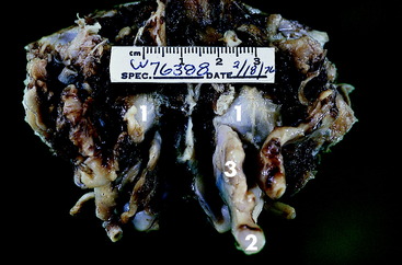
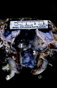
Figure 21-2 Dorsal view of the caudal cranial fossa of the skull in Fig. 21-1. The neoplasm (1) on the left is continuous through the jugular foramen and tympanooccipital fissure with the enlarged vagosympathetic trunk (2) and associated ganglia seen in Fig. 21-1. Note the normal jugular foramen (3) with branches of cranial nerves IX, X, and XI in it.
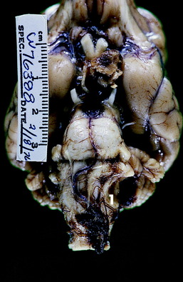


The course of the clinical signs, with the sympathetic and pharyngeal nerves being the first presumed to be involved, is highly suggestive that the neoplasm began outside the cranial cavity in the region of the tympanooccipital fissure. This suggestion is further supported by the late onset of the vestibular system dysfunction. No indication was found as to when the hypoglossal involvement began because this information would have required direct visualization of the hemiatrophy of the tongue. The involvement of the intracranial portion of the accessory nerve, the extracranial portion of the vagus nerve, or both may also have caused a left laryngeal hemiparesis, which might have contributed to the coughing seen initially.


Remember that Horner syndrome can be caused by lesions in a wide range of anatomic locations. The presence of other clinical signs of neurologic dysfunction usually determines the site of the lesion in the sympathetic system. Dogs with persistent choking, gagging, or coughing for which no explanation can be found in the pharynx should be evaluated for a possible partial paralysis of the pharyngeal muscles. If paralysis is suspected, remember to palpate the area of the tympanooccipital fissure through the dorsolateral wall of the laryngopharynx via the mouth. The mass in this dog may have been palpated antemortem if paralysis had been suspected. CT or MR imaging would facilitate detection of this mass lesion but would not likely have altered the long-term outcome.


CASE EXAMPLE 21-2


Signalment: 11-year-old female collie


Chief Complaint: Inability to use the pelvic limbs


History: Ten days before the examination, this dog became lame in the left thoracic limb. One week later, she began to drag the left pelvic limb, and within 3 days, she became recumbent in the pelvic limbs and unable to stand up with either pelvic limb.


Examination: This collie had been recumbent for the entire day before this examination. She lay in lateral recumbency and was unable to assume a sternal position. When held up and supported by the trunk and tail, she walked on the thoracic limbs with short stiff strides. She was paraplegic (grade 0) and dragged the motionless stiff pelvic limbs. The trunk had to be supported, otherwise she would sway to the side and fall. No postural reactions were observed in the pelvic limbs. The right thoracic limb was stiff but hopped normally. The left thoracic limb hopped slowly with a mild delay in the onset of protraction. Both pelvic limbs and the right thoracic limb were hypertonic. The left thoracic limb was hypotonic, and most of its muscles were mildly atrophied. Both patellar reflexes were brisk (+3). The flexor reflexes were normal in all four limbs. Nociception was depressed in the pelvic limbs (hypalgesia) but normal in the thoracic limbs. Testing for nociception along the trunk revealed a suspicious line in the cranial thoracic region with hypalgesia caudal to it. On cranial nerve examination, the only abnormalities involved the left orbit, where slight miosis, protrusion of the third eyelid, and a smaller palpebral fissure were seen.


Anatomic Diagnosis: Cranial thoracic spinal cord lesion that is functionally transverse at T3 and extends cranially on the left side into the cervical intumescence. The nearly transverse lesion at T3 is the cause of the paraplegia and the loss of trunk strength (UMN paresis), causing it to sway unless supported. Partial involvement of the left side of the T2 and T1 spinal cord segments or ventral roots along with the T3 lesion is the cause of the left-side Horner syndrome. Partial involvement of the left side of the T1 and C8 spinal cord segments or ventral roots is the cause of the mild left thoracic limb deficit and muscle atrophy.


Differential Diagnosis: Neoplasm—nerve sheath, meningioma, metastatic, glioma; intervertebral disk extrusion-protrusion; inflammation


The localizing clinical signs, their asymmetry, and the progressive course are all supportive of a presumptive diagnosis of an extramedullary neoplasm compressing the spinal cord. Extramedullary neoplasms are more common than intramedullary neoplasms and are more likely to cause asymmetric clinical signs as observed here.


Nerve sheath neoplasms commonly affect spinal nerves and occasionally the sympathetic trunk and often extend into the vertebral canal and compress the spinal cord. If the sympathetic trunk were involved with a nerve sheath tumor at the level of the cervicothoracic ganglion, Horner syndrome would have preceded the clinical signs of spinal cord involvement. A vertebral body or epidural metastasis at this site might also explain the clinical signs. Extramedullary neoplasms may grow slowly to substantial size and slowly compress the spinal cord without causing any clinical signs. The amount of compression that can occur is quite extraordinary, as long as the process is very slow. However, a critical point apparently exists in the ability of the CNS autoregulation that maintains normal spinal cord perfusion to function, and when this limit is surpassed, ischemia occurs with an abrupt onset of clinical signs that rapidly progress. Anesthesia for ancillary studies of these patients may precipitate the loss of autoregulation and result in the presence of more severe clinical signs after recovery from the anesthesia.


Intervertebral disk extrusion-protrusion is uncommon in the cranial thoracic vertebral column and would be less likely to cause the asymmetric thoracic limb clinical signs. Of the various inflammations that affect the spinal cord, focal granulomatous meningomyelitis, or a focal fungal myelitis would be the most likely candidates. Most viral or rickettsial infections cause multifocal or diffuse lesions. Vascular compromise such as fibrocartilaginous embolic myelopathy (FCEM) was not considered because of the progressive nature of the clinical signs.


Ancillary Procedures: Radiographs were normal. A lumbar myelogram showed the contrast to stop abruptly at the caudal aspect of the body of T2, where a small amount of contrast escaped from the subarachnoid space, and the vertebral canal to appear dorsally adjacent to the spine of T3 and ventrally along the ventral aspect of the vertebral body of T2. The lumbar CSF contained no leucocytes and 31 mg/dl of protein (normal <25). These findings were considered to be supportive of the presumptive diagnosis of an extramedullary neoplasm and compressive myelopathy.


Outcome: Exploratory surgery was recommended. However, based on the age of the dog and the severity of the clinical signs, the owner elected euthanasia.


At necropsy, a large firm nodular mass was found in the thorax on the surface of the left longus colli muscle involving the cranial aspect of the thoracic sympathetic trunk and left cervicothoracic ganglion. The mass extended dorsally between the first two ribs and entered the vertebral canal through the intervertebral foramen between the first and second thoracic vertebrae. It compressed the left first thoracic spinal nerve and expanded into the epidural space where it compressed the spinal cord to the right side (Fig. 21-4). The second and especially the third spinal cord segments were the most compressed. The third thoracic spinal cord segment was compressed to less than one half of its normal width. On microscopic examination this neoplasm was diagnosed as a malignant nerve sheath neoplasm.


This case emphasizes the importance of doing a complete neurologic examination regardless of the chief complaint. Recognition of Horner syndrome was especially of value in localizing the level of the lesion in the spinal cord of this dog. This syndrome probably occurred before the gait deficit if it can be assumed that the sympathetic trunk and cervicothoracic ganglion were affected by the neoplasm initially before the neoplasm extended into the vertebral canal. Most extramedullary spinal cord neoplasms are good candidates for surgical removal. However, recurrence is common for this nerve sheath neoplasm, given that removing all the neoplastic cells is impossible. Surgery or radiation therapy would not likely have significantly altered the long-term outcome in this dog given the location of the lesion in the thoracic sympathetic trunk and the relatively aggressive nature of this particular neoplasm. These types of neoplasms are readily identified by CT or MR imaging. See Video 7-2 and Case Example 7-1 for a similar disorder.


CASE EXAMPLE 21-3


Signalment: 8-year-old male coonhound mixed breed


Chief Complaint: Unable to stand up


History: Approximately 4 weeks before the examination, this dog was noticed to be slow on treeing a raccoon. This slow reaction occurred 1 month after the dog had been bitten by a raccoon. The dog continued to slow down when hunting and then exhibited a persistent abnormal gait that progressively worsened. Six days before the examination, the dog became recumbent and was unable to stand up without assistance.


Examination: See Video 8-1. This dog was considered to be mildly depressed. He was unable to stand unassisted. When held up, he veered off to the right side and made slow walking attempts with stiff awkward movements as he leaned to the right side. The limbs were markedly hypertonic (spastic), and his ability to support weight was unimpaired. However, he acted very disoriented. The head was tilted to the right, and the neck was curved, with the concavity on the right side (pleurothotonus). He would occasionally exhibit an opisthotonic head and neck posture with the thoracic limbs extended and the pelvic limbs extended but usually flexed at the hips. The trunk often weaved from side to side, and a mild head tremor was evident.


The dog had to be held up to test the postural reactions. The hopping responses were slow in all four limbs but were slower in the left limbs. He fell when hemiwalking was attempted with the left limbs. Paw replacement was absent with the left paws.


He preferred to lie in left lateral recumbency. All the limbs were hypertonic, but hypertonia was worse in the left limbs when he was placed in right lateral recumbency. No muscle atrophy was observed. Patellar reflexes were brisk (+3). All flexor reflexes were normal, as was the perineal reflex. Nociception was normal.


On cranial nerve examination, the only abnormalities were referable to the vestibular system. A right head tilt was seen. On neck extension, a rotatory right positional nystagmus was observed, and the right eyeball did not elevate fully in the palpebral fissure. No strabismus was seen in other head positions, and physiologic nystagmus was normal.


Anatomic Diagnosis: Caudal cranial fossa—cerebellum, pons, and medulla


Despite the breed and use of this dog and the observed exposure to raccoon bites, these clinical signs are the antithesis of the clinical signs of polyradiculoneuritis of coonhound paralysis, which are diffuse neuromuscular signs. The vestibular system dysfunction was severe with the head tilt, the leaning to one side, the abnormal nystagmus, and the strabismus. The spastic tetraparesis and general proprioceptive (GP) ataxia and decerebellate posture clearly indicate that the clinical signs of vestibular system dysfunction are caused by involvement of its central components. Note that the direction of the abnormal nystagmus also supports a central lesion in this system. The decerebellate posture implicates dysfunction of the rostral portion of the cerebellum. The spastic tetraparesis and GP ataxia support involvement of the pontomedullary UMN and GP systems, with the left side more affected. At this level, the UMN system is primarily ipsilateral to the limbs it controls. The portion of the GP system primarily involved here on the left side would be the spinocerebellar tracts, which are also primarily conducting sensory information from the ipsilateral left limbs. The loss of the left medial lemniscus may contribute to the right limb deficits. The predominance of right-sided central vestibular system clinical signs may reflect an asymmetric bilateral caudal cranial fossa lesion or a multifocal lesion, or these may be paradoxical vestibular clinical signs from a left-side lesion involving the left cerebellar peduncles.


Differential Diagnosis: Neoplasm, focal inflammation, epidermoid or dermoid cyst


The 1 month of slowly progressive clinical signs with a rapid deterioration in the last week supports a presumptive diagnosis of an extramedullary neoplasm such as a meningioma, choroid plexus papilloma, multilobular bone tumor, or some form of sarcoma. These extramedullary neoplasms at this location commonly cause a slow progression of clinical signs. An intramedullary neurectodermal tumor is a strong consideration but only slightly less likely as a result of the prolonged progressive history. Any possibility of a progressive caudal cranial fossa masslike lesion must include an abscess from an intracranial extension of an otitis media-interna. Focal GME occasionally occurs in the cerebellar white matter with involvement of the adjacent pons and medulla. A fungal granuloma can occur at this level of the CNS. The protozoal infections must be considered. Toxoplasmosis is not common and is usually multifocal, but it might cause a large granuloma at this level. Neospora caninum is noted for causing an extensive chronic inflammation in adult dogs that can be remarkably limited to the cerebellum. The viral and rickettsial infections are less likely to produce such severe focal caudal cranial fossa clinical signs. Although epidermoid and dermoid cysts are most common in the caudal cranial fossa, the age of this patient and the severity of the clinical signs make this consideration less likely. The progressive course of this disorder eliminates any consideration of a vascular compromise despite the risk of a cerebellar infarction in older dogs. The clinical signs were too focal, asymmetric, and severe to give any consideration for an inherited degenerative disorder.


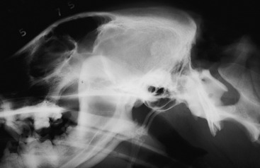
Ancillary Studies: Radiographs revealed an oval, hyperdense lesion that engulfed the tentorium cerebelli (Fig. 21-5). A meningioma or sarcoma was suspected to be the cause of this hyperdense mass lesion. Cisternal CSF on day 2 of hospitalization contained 14 mononuclear cells/mm3 (normal <5) and 98 mg/dl of protein (normal <25). A second cisternal CSF obtained on day 4 of hospitalization contained 40 white blood cells (WBCs)/mm3, most of which were mononuclear cells, and 176 mg/dl protein. The CSF opening pressure was 265 mm water (normal <180). These ancillary results were compatible with an extramedullary neoplasm involving the tentorium cerebelli, causing compression of the cerebellum and caudal brainstem. Be aware that obtaining CSF from a dog with an intracranial space-occupying lesion has the risk of possible brain herniation secondary to increased intracranial pressure. Today, a delay in obtaining CSF until after advanced imaging has determined the nature of the brain lesion is usually standard procedure.


Outcome: On the second day of hospitalization, all the clinical signs were slightly worse. In addition, the left pupil was slightly dilated but still responsive to light. Occasionally, dysfunction of the cerebellar nuclei will cause an anisocoria and may therefore be the result of the cerebellar compression in this dog or the result of a direct compression of the left side of the mesencephalon, with partial dysfunction of the parasympathetic general visceral efferent neurons of the oculomotor nerve. On day 4 of hospitalization, the patient’s clinical signs of decerebellation were worse, and he made fewer attempts to stand. Surgery to remove the neoplasm was recommended, but the owners declined and elected euthanasia.


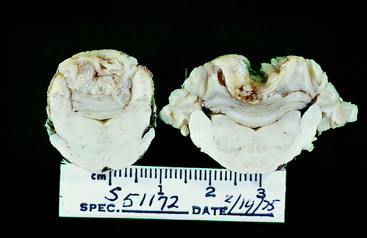
Necropsy confirmed the presence of a large very firm nodular mass that enveloped the tentorium cerebelli (Fig. 21-6) and primarily projected ventrally where it severely compressed the rostral and central portions of the cerebellum (Figs. 21-7,21-8). At the most compressed level of the cerebellum, it only measured 7 mm in height. The caudal cerebellar vermis projected caudally into the foramen magnum. The entire pons and medulla were compressed by the adjacent cerebellar compression. Dorsal to the tentorium cerebelli, the neoplasm compressed the medial portions of both occipital lobes. On microscopic examination, the neoplasm was diagnosed as an osteogenic sarcoma.


image

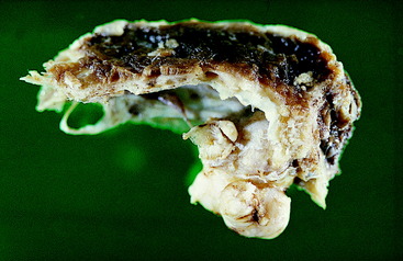
Figure 21-6 Lateral view of the preserved calvaria from the skull of the dog in Fig. 21-5, showing an osteogenic sarcoma involving the tentorium cerebelli.


image

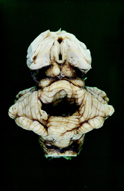
Figure 21-7 Dorsal view of the preserved brainstem and cerebellum of the dog in Figs. 21-5 and 21-6. Note the deep depression in the center of the cerebellum where the tentorial neoplasm was removed with the calvaria.


image

Figure 21-8 Transverse sections of the preserved caudal brainstem and cerebellum of the dog in Figs. 21-5,21-6, and 21-7, showing the depression in the cerebellum and compression of the pons and medulla caused by the tentorial osteogenic sarcoma.


CASE EXAMPLE 21-4


Signalment: 3-year-old female setter mixed breed


Chief Complaint: Abnormal pelvic limb function


History: Five days before examination, this dog ran off from its home. She was found 4 days later lying alongside the road and unable to stand up and exhibiting extreme discomfort. She tried to bite anyone who attempted to move her.


Examination: She was alert and responsive and stood up on her thoracic limbs but refused any attempts to stand on her pelvic limbs. When assisted to a standing position, she walked normally on the thoracic limbs, supported her trunk well, and supported her weight on her right pelvic limb, which she moved slowly but without any obvious neurologic deficit. The dog was reluctant to stand on her left pelvic limb but was able to bear weight if positioned carefully. She often stood on the dorsal surface of her left paw and could move the limb only by mild hip flexion.


Muscle tone, spinal nerve reflexes, and nociception were all normal in the right pelvic limb. The left pelvic limb was mildly hypotonic. The patellar reflex was normal. No flexor reflex was observed when a noxious stimulus was applied to digits 3, 4, and 5, and nociception was absent from these digits. Analgesia was observed from all aspects of the paw except for the medial side. A noxious stimulus to the skin on the medial aspect of the limb at any level resulted in slight hip flexion and intact nociception but no flexion of the stifle, tarsus, or digits. Cutaneous analgesia was observed in the caudal thigh, as well as all of the crus, tarsus, and paw, except for the medial side of the entire left limb. The tail was atonic, areflexic, and analgesic. The anus was hypotonic and partially dilated. A slight anal closure response to stimulation of the perineum was seen on the right side but none from the left side. Nociception was intact on the right side of the perineum but the left side was analgesic. The bladder was distended and required manual evacuation. In addition to these neurologic deficits was swelling at the base of the tail, and crepitus was palpated. The normal space between the right greater trochanter and the right tuber ischium was enlarged.


Anatomic Diagnosis: Left sciatic nerve or L6, L7, and S1 spinal nerve ventral branches, spinal nerves, spinal nerve roots, or spinal cord segments; branches of the left sacral plexus, sacral spinal nerves, spinal nerve roots and spinal cord segments; bilateral pelvic nerve and caudal spinal nerve branches, spinal nerve roots, and spinal cord segments


The femoral nerve and its L4 and L5 spinal cord and spinal nerve components were normal based on the normal patellar reflexes and normal nociception from the medial side of the paw and crus via the saphenous nerve branch of the femoral nerve. The normal cutaneous sensation over the cranial thigh resulted from the unaffected lateral cutaneous femoral nerve (L3 and L4) and over the proximal medial thigh from the intact genitofemoral nerve (L3 and L4). The normal hip flexion resulted from the intact innervation of the psoas major muscle from the ventral branches of most of the lumbar spinal nerves. The bladder paralysis requires a bilateral loss of the pelvic nerve innervation (S1, S2, and S3) The loss of the left perineal reflex, nociception, and anal tone requires dysfunction of the left pudendal nerve or its sacral plexus and nerve origin on the left side. The tail deficit requires a bilateral loss of caudal spinal cord segment or spinal nerve innervation.


Differential Diagnosis: The nature of the onset of the clinical signs is unknown because of the lack of observation of the dog during that time period. Finding the dog beside the road in significant discomfort with palpable skeletal abnormalities in the area of the pelvis is strongly presumptive of a traumatic event of external origin. Most likely this dog was struck by a vehicle, and the clinical signs are the result of injury, primarily of the peripheral nerve components of the structures listed in the anatomic diagnosis. The sciatic nerve or its spinal nerve branches of origin are commonly injured by fractures of the ilium or ischium or sacroiliac luxations because of their close anatomic relationship. Sacrocaudal nerves are commonly injured by sacrocaudal vertebral fractures. Pelvic nerve dysfunction can result from hemorrhage in the connective tissues of the pelvic cavity.


Ancillary Studies: Radiographs revealed a right coxofemoral joint luxation with a craniodorsal dislocation of the right femur, a fracture of the sacrum at the left sacroiliac joint with subluxation, and a fracture of the sacrocaudal articulation with complete ventral displacement of the caudal vertebral portion (Figs. 21-9,21-10). The ventral branches of the L6 and L7 spinal nerves that contribute to the formation of the sciatic nerve course caudally on the ventral surface of the sacroiliac joint to be joined by the ventral branches of the first and second sacral spinal nerves to form the sciatic nerve at the level of the greater ischiatic notch of the ilium. The skeletal lesions seen here correlate well with the anatomic diagnosis and the clinical signs observed in this dog. Remember that your neurologic examination determines a functional abnormality in the nervous system and not the structural basis for it. Therefore be cautious about your prognosis, especially with injuries such as this one.


image

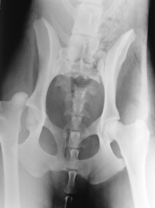
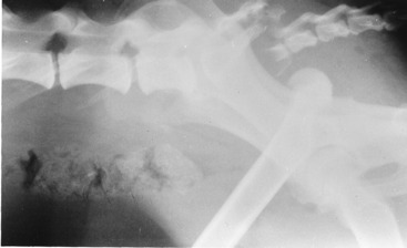
Figure 21-10 Lateral radiograph of the pelvis of the dog in Fig. 21-9, showing the sacrocaudal fracture and the luxated right femur. Note the asymmetry of the ilia caused by the subluxation of the left sacroiliac joint.


Outcome: The right coxofemoral luxation was manually replaced under general anesthesia. Within a few days, this dog recovered its sensation in the distribution of the left sciatic nerve, but motor function remained absent. The right pelvic limb was normal in all functional respects. Within the following 10 days, the dog began to stand and make slow attempts at walking with the left pelvic limb. She supported her weight well (normal femoral nerve) and would advance the left pelvic limb by hip flexion (normal iliopsoas muscle and lumbar spinal nerves). However, she walked with the tarsus overflexed and the stifle extended and often placed the paw on its dorsal surface on the ground. The tarsus was lax from loss of muscle tone. These clinical signs were caused by the persistent sciatic nerve paralysis. Incontinence persisted from the pelvic nerve injury, and the tail remained paralyzed and analgesic. After 1 more month with no further improvement, the owners requested euthanasia for their dog.


At necropsy the skeletal lesions were observed that were diagnosed on the radiographs. The caudal spinal nerves were torn and fibrosed at the sacrocaudal fracture. The left L7 spinal nerve ventral branch was discolored and entirely embedded in fibrous tissue ventromedial to the left sacroiliac joint subluxation. A portion of the left L6 spinal nerve ventral branch was involved in the same fibrous adhesion. The ventral branches of the sacral spinal nerves were embedded in old hemorrhage and fibrous tissue. No spinal cord lesions were observed.


CASE EXAMPLE 21-5


Signalment: 8-year-old female terrier mixed breed


Chief Complaint: Head tilt


History: Six weeks before examination, this dog rapidly developed a mild right head tilt. The degree of head tilt slowly worsened, and for the past 10 days, she tended to move to her right as she walked.


Examination: The patient was alert and responsive. Her head was held markedly tilted to her right side. Her gait was normal except for a tendency to drift to the right side.


Hopping responses were slow in the right limbs. Muscle size and tone and all spinal nerve reflexes were normal. Nociception was normal. Cranial nerve examination revealed anisocoria caused by a slightly enlarged right pupil. Menace responses and pupillary light reflexes were normal, except that the right pupil never would constrict as much as the left pupil. The right eyeball was frequently held in a ventrolateral position but did not persist in all head positions. On testing physiologic nystagmus by moving the head back and forth, both eyeballs adducted and abducted fully. No spontaneous resting nystagmus was observed, but a positional rotatory left-to-vertical nystagmus developed when the head was held flexed to the left side.


Anatomic Diagnosis: Two considerations: (1) focal right side caudal cranial fossa lesion—pons, medulla, cerebellum and (2) focal right middle- and inner-ear plus left prosencephalon


Stay updated, free articles. Join our Telegram channel

Aug 26, 2016 | Posted by in INTERNAL MEDICINE | Comments Off on Case Descriptions

Full access? Get Clinical Tree

Get Clinical Tree app for offline access