Cardiomyopathy

Chapter 150 Cardiomyopathy


OVERVIEW


The left and right ventricles are capable of undergoing significant morphologic change in response to stresses and stimuli. Reaction to increased volume or pressure work frequently leads to chamber dilatation and hypertrophy, along with alterations in the cytoskeletal matrix of the ventricle. Myocardial and chamber responses develop with chronic valvular heart disease, cardiac shunts, systemic and pulmonary hypertension, thyrotoxicosis, and chronic anemia. These responses to stress are directed by various neural, hormonal, and genetic messages acting on the myocardium. These signals influence genetic expression in the cardiomyocyte and interstitial cells, and remodel the ventricle.


“Cardiomyopathy” refers to disease of the myocardium and, by extension, the cardiomyocyte and the supporting collagen and interstitial matrix. Idiopathic or primary cardiomyopathies are those that cannot be explained by a malformation, acquired cardiac lesion, dysrhythmia, or coronary artery disease. Many idiopathic cardiomyopathies are genetic diseases. A more expansive definition of cardiomyopathy accepts that some myocardial diseases can be explained by other disorders, and in such cases, the term secondary cardiomyopathy can be used. When myocardial failure develops from chronic volume or pressure overload of the ventricle, the term cardiomyopathy of overload has been proposed to explain the remodeling associated with increased ventricular work.


Although there are many known causes of cardiomyopathy, most cases in cats and dogs are idiopathic and thought to represent a genetic disorder. This is particularly true of dilated cardiomyopathy in dogs and hypertrophic cardiomyopathy in cats. What stimulates these genetic factors to cause heart muscle disease is poorly understood, and most cases of cardiomyopathy are irreversible and progressive. But there are special examples that demonstrate that some cardiomyopathic states can be postponed, arrested, or even reversed. For example:


Regression of left ventricular hypertrophy may occur after successful treatment of systemic hypertension or hyperthyroidism.

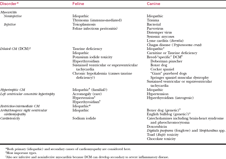
Cardiomyopathies often are classified by the postmortem anatomic appearance of the left (or right) ventricle and by the correlative echocardiographic features of ventricular anatomy and function. The most important forms of cardiomyopathy can be classified as follows (see Table 150-1):


Cardiotoxicity—The heart also can be damaged by a number of cardiotoxins, some of which are listed in Table 150-1. The outcome of cardiotoxicity is often an arrhythmia, conduction disturbance, sudden death, or development of a secondary dilated cardiomyopathy.


This chapter will next describe the clinical features of feline cardiomyopathies and the therapy of related complications. Following this is a consideration of canine DCM and arrhythmogenic cardiomyopathy.


FELINE HYPERTROPHIC CARDIOMYOPATHY


Overview and Pathophysiology of Feline HCM


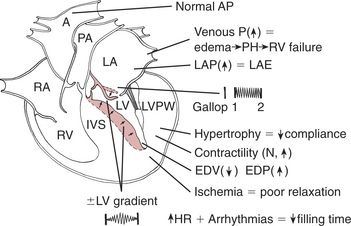
The pathophysiology of feline HCM (Figure 150-1) is relevant to the diagnosis and drug therapy of this disease. The presumptive cause of CHF is ventricular diastolic dysfunction, because most cats with HCM have a normal to hyperdynamic LV ejection fraction.


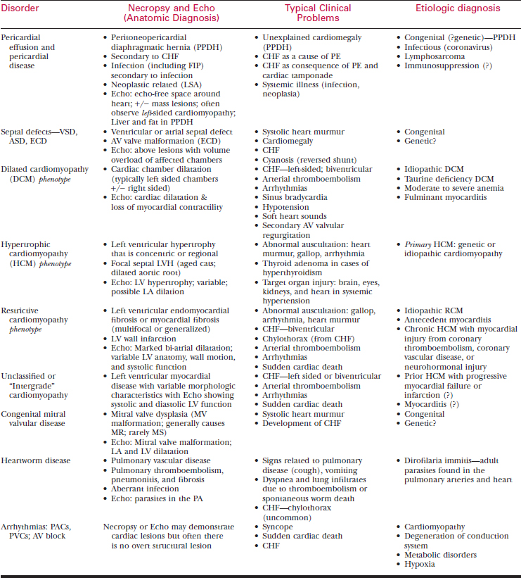
Table 150-2 DIFFERENTIAL DIAGNOSIS OF FELINE CARDIOMYOPATHY AND HEART FAILURE*


Clinical Findings in Feline HCM


The clinical presentation and examination findings in feline HCM are variable.


Diagnostic Tests in HCM


A number of routine diagnostic tests are helpful in recognizing and staging HCM.


Echocardiography—Definitive diagnosis and staging of HCM relies on echocardiography and Doppler studies interpreted in light of clinical findings.


The differential diagnosis of feline cardiomyopathy is extensive (Table 150-2), including other cardiovascular and noncardiac disorders.


FELINE RESTRICTIVE CARDIOMYOPATHY (RCM)


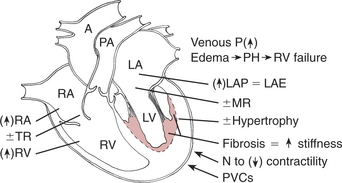
Feline RCM represents a heterogeneous disorder, and some latitude is used in placing cats within this category. The disorder described below might be interpreted by others as “intermediate cardiomyopathy” or as “unclassified cardiomyopathy.”


Overview and Pathophysiology of Feline RCM


Clinical Findings in Feline RCM


DILATED CARDIOMYOPATHY IN CATS


As shown by Pion and colleagues, dietary deficiency of taurine accounted for the vast majority of feline cases of dilated cardiomyopathy. Today DCM in cats is a relatively rare occurrence requiring echocardiographic evaluation for diagnosis. Taurine deficiency may still be observed in cats eating mostly “custom” diets or dog food, but most cases of DCM are idiopathic or a consequence of myocarditis.


OTHER FELINE MYOCARDIAL DISEASES


A number of other diseases that affect the myocardium of the cat are briefly considered below.


Aug 27, 2016 | Posted by in SMALL ANIMAL | Comments Off on Cardiomyopathy

Full access? Get Clinical Tree

Get Clinical Tree app for offline access