Canine Infectious Diseases

69 Canine Infectious Diseases


SUMMARY OF DIAGNOSTIC TECHNIQUES


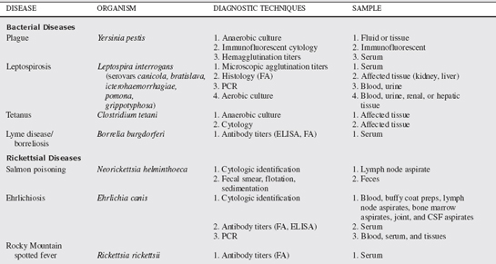
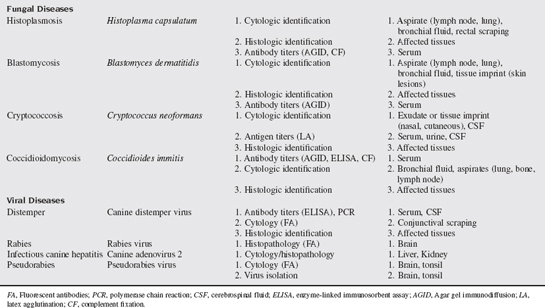
A multitude of infectious diseases are encountered in small animal veterinary practice and, as such, numerous diagnostic tests and techniques are used in their diagnosis. Some of the commonly used techniques and tests available in small animal infectious disease diagnosis are discussed in this chapter. Specific tests for the diseases discussed in this chapter are summarized in Table 69-1.


1. How are antigens and antibodies detected?

Serologic testing refers to the detection of endogenous antibodies directed against a particular infectious agent or detection of actual infectious antigens. Numerous techniques are used in this process. Serologic antigen detection is considered more diagnostic of infection than antibody detection because antibodies can be present in animals that were previously infected. Additionally, serologic antibody tests are more likely to have cross-reactivity with antibodies directed at other, closely related agents. Therefore high single antibody titers, or demonstration of a fourfold increase in serum antibody titers, are generally required to support active infection.


The enzyme linked immunosorbent assay (ELISA) is a commonly used rapid method of detection of antibodies or antigens. This methodology is used in commercial test kits intended for “in-house” use. Dirofilaria (antigen), Ehrlichia (antibody), and Borrelia (recombinant product that detects antibody) infections can be detected in practice settings with these kits. Individual test characteristics are important for the practitioner to understand, because false-positive and false-negative tests results are not uncommon when there is a lack of quality control in their performance.


Fluorescent antibodies (FAs) can also be used to detect antibodies or antigen. These are not performed as an in-house test. A fluorescing molecule is first bound to antibodies specific to an organism or antibody being evaluated. These are then applied to serologic, histologic, or cytologic specimens. If the organism or antibody of interest is present in the sample, fluorescence will be seen when evaluated. Fluorescent techniques have some subjectivity to their interpretation and the practitioner should be confident of individual laboratories performing these interpretations. Diseases diagnosed with this methodology include distemper, rabies, pseudorabies, Lyme disease, Rocky Mountain spotted fever (RMSF), ehrlichiosis, plague, and tularemia.


The ability of antigen or antibody to cause agglutination of red cells or other materials is used for detection of some infectious diseases. Latex agglutination is used in a kit for cryptococcal antigen detection. Agar gel immunodiffusion (AGID) and complement fixation (CF) are used to detect antibodies against some of the pathogenic fungi. In general, these two tests lack sensitivity and specificity and therefore identification of the organism is important.


The availability of polymerase chain reaction (PCR) as a method of detection is gradually increasing in veterinary medicine. Minute amounts of DNA or RNA can be detected with nucleic acid primers specific to the organism and then multiplied via rounds of replication to produce enough material for electrophoretic analysis. As a result, PCR tests can detect infections in tissues with low concentrations of antigen and detect differences in species within a genus. Along with the incredible sensitivity of these tests comes a greater effect on results if contamination occurs. Whole blood can be collected from dogs and tested via PCR for Bartonella and Ehrlichia species. Blood or joint fluid can be tested for Borrelia. Urine or renal tissue can be tested for Leptospira. Other PCR tests for canine infectious diseases are likely to become commercially available in the near future.


2. How do you collect cytologic samples?

Lymph node and tissue aspiration are simple, inexpensive, and frequently rewarding techniques used to diagnose cases of fungal infection. A 22-gauge needle is applied to a 12-cc syringe. The needle is then inserted into the affected tissue, and either it is redirected through the tissue numerous times (“woodpecker” technique), or a light amount of suction is applied by drawing back to the 2- or 3-cc mark on the syringe. Suction is released and the needle withdrawn. The needle is removed from the syringe, air drawn into the syringe, the needle reattached, and the fluid forced out of the syringe onto a slide, culture swab, or into a glass tube depending on the intended purpose of the sample. In the case of cytology, a simple smear is made. Cytologic samples can be used for direct visualization of Histoplasma, Blastomyces, Coccidioides, Cryptococcus, Neorickettsia helminthoeca, Ehrlichia, and Clostridium. With application of FA techniques, cytologic samples can be used to verify Yersinia and Tularemia infections.


Conjunctival scrapings can be used for cytologic diagnosis of distemper. Scrapings are collected by first removing mucus and tears from the inferior conjunctiva with a cotton tip applicator. A heat-sterilized blunt metal spatula or dull scalpel blade is then used to repeatedly scrape the superficial layer of conjunctiva until a small amount of tissue can be lifted off, applied to a glass slide, and smeared. This usually causes erythema of the conjunctiva and a slight amount of bleeding. Slides are sent for distemper fluorescent antibody staining. Using routine in house stains, the slides can be evaluated for characteristic intracytoplasmic distemper virus inclusions.


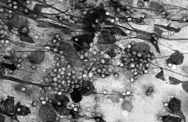
Rectal scrapings can be useful for detection of histoplasmosis (Fig. 69-1). This is performed by passing a gloved finger or cotton-tipped applicator into the rectum and scraping the mucosa repeatedly. The finger or applicator is removed and the tip lightly rolled on a slide for staining with routine in house stains.


Tracheal washes can be used for diagnosis of fungal and bacterial infections. A 16- to 18-gauge, 12-inch through-the-needle catheter is needed. The needle is either inserted between tracheal rings either in the distal third of the cervical trachea large dogs or in the cricothyroid ligament (space just cranial to the firm ventral ridge of the cricoid cartilage) in medium-size dogs. The area of puncture is prepared aseptically and sterile gloves are worn. Dogs that weigh less than 20 pounds are best sampled by passing a sterile red rubber catheter through a sterile endotracheal tube. After the needle is inserted and the catheter advance through it, the needle is withdrawn a short distance out of the skin. One to 10 ml of sterile saline is infused. When the animal coughs, aspirate to collect wash material. This can be repeated several times if needed. The catheter is removed and typically no bandaging is needed. Subcutaneous emphysema can occur. The fluid is used for cytologic analysis and culture as indicated.


3. How do you collect and evaluate cerebrospinal fluid (CSF)?

CSF is collected in a much more refined manner and must be handled with care. Cytologic preparations are best interpreted by an experienced cytologist and made after cytocentrifugation to concentrate the low quantity of cells into a small area. The fluid must be analyzed within a short period before cellular degradation. Therefore a laboratory capable of handling CSF should be identified before collection and be reachable within 30 minutes. The atlantooccipital cistern is the most useful site of collection for most central nervous system (CNS) infections. After anesthesia has been induced and the site aseptically prepared, the dog is placed in lateral recumbency (right lateral for a right dominant person) with the neck flexed to open the atlantooccipital cistern. The space is identified as a subtle depression in the center of a triangle made by placing the left thumb on the right wing of the atlas, the left middle finger on the left wing, and the index finger on the occipital protuberance. This correlates to the cranial aspect of the atlas. A 22-gauge, 1.5- to 2.5-inch spinal needle is then slowly inserted into this space with the needle nearly 90° to the spine and directly on midline with the bevel directed cranially. It is uncommon to completely insert a 1.5-inch needle into a dog of any size. The stylet is removed intermittently to evaluate for flow so that the needle is not advanced into the cord or brain stem. If frank blood appears, the needle has been placed off midline and the needle should be removed. After CSF flows from the needle, it should be aseptically collected by allowing it to drip into an empty glass tube. Approximately 1 to 2 ml is required for most diagnostic tests. The needle is then removed. Cell counts, cytologic evaluation, and fluorescent antibody techniques can be applied to CSF for detection of canine distemper virus antigens. Titers for distemper, Ehrlichia canis, RMSF, Cryptococcus, and other infections can be determined and compared with serum concentrations for diagnosis. Fluid can be submitted for cultures.


VIRAL DISEASES


Canine Distemper


7. What are the major clinical manifestations of canine distemper?

Major clinical signs develop in young animals usually younger than 16 weeks. The most common systems affected by canine distemper include respiratory, gastrointestinal, skin and nervous systems.


Ocular and nasal discharges are present in early stages of the disease, and are usually associated with concomitant constitutional signs such as fever, lethargy, and anorexia. If the dog’s immunity is inadequate, secondary bacterial infections result in purulent ocular and nasal discharges and cough. Respiratory distress may be observed in severely infected animals.


Gastrointestinal signs such as vomiting and diarrhea may be observed because of the local replication of virus in these tissues. Enamel hypoplasia can be noted in young animals that were previously infected with canine distemper and subsequently develop neurologic signs.


Dermatologic signs such as impetiginous dermatitis in young dogs, or nasal and footpad hyperkeratosis may be noted. Pustular dermatitis is reported to be a good prognostic indicator but is not a consistent observation.


Neurologic signs in dogs are due to acute or chronic encephalitis. Manifestations include seizures, cerebellar and vestibular signs, paraparesis, tetraparesis, and myoclonus. Myoclonus and the classical “chewing gum fits” are features that are almost pathognomonic for this disease, but other CNS disease can produce similar clinical manifestations. However, if these signs are observed in a susceptible, unvaccinated young dog, it would lend more credence to canine distemper infection. Ocular manifestations associated with canine distemper infection include conjunctivitis, keratitis, optic neuritis, and keratoconjunctivitis sicca.


9. What are major means of diagnosis of CDV infection?

A compatible history and clinical signs are usually supportive of a diagnosis of acute canine distemper. Lack of proper vaccination in a young animal should also increase the clinical suspicion. Chronic neurologic disease from canine distemper infection is difficult to differentiate from other causes of acquired seizures and paresis, based solely on historical and physical findings.


CSF analysis in dogs with acute encephalitis usually shows increases in protein concentration and increased mononuclear cells, predominately lymphocytes. Detection of anti-CDV antibody in the CSF is the most definitive evidence of distemper encephalitis, provided that blood contamination is minimal during collection. If blood contamination is present in CSF, serum, and CSF antibody titers for CDV and canine parvovirus can be measured and their corresponding CSF to serum ratios computed. If the ratio for CDV antibodies exceeds that of parvovirus, then a diagnosis of CDV infection is plausible (see Chapter 4). Immunocytology and immunohistochemistry can be used to detect viral antigen in tissue samples. Conjunctival, tonsillar, and respiratory samples may reveal a positive fluorescence test early in the course of disease before antibody titers increase. This technique may also be applied to tissue samples obtained at the time of biopsy or necropsy.


Serum titers can be detected by ELISA methodology. Immunoglobulin (Ig)M antibodies can be detected in both acute and chronic encephalopathies from canine distemper and is more definitive than IgG antibodies. IgG antibodies only denote that the animal has been exposed to the virus, either naturally or through vaccination.


A seminested reverse transcriptase PCR has also been used to detect viral antigen in fresh and formalin fixed tissues to aid in the detection of canine distemper.


Canine Adenovirus Infections


17. What are the major clinical manifestations of adenovirus infections in dogs?

CAV-1 infections can be commonly asymptomatic infections, based on serologic surveys, or cause acute or chronic disease syndromes. These syndromes are related to organ injuries of the liver, kidneys, eyes, and respiratory tract. The predominant organ injury with CAV-1 is the liver, where it causes hepatic necrosis. Clinical signs related to hepatic injury include fever, vomiting, diarrhea, hepatic enlargement, and abdominal pain. If the dog mounts an effective immune response early in the disease, hepatic injury may be limited. In dogs with severe hepatic necrosis, disseminated intravascular coagulation (DIC) may result and cause a hemorrhagic diathesis. Partial immune responses in infected CAV-1–infected dogs can result in a chronic hepatopathy, characterized by persistent hepatic inflammation.


Renal injury also occurs in CAV-1 infection but is subclinical. The virus localizes in the glomerular vasculature and later leads to immune complex deposition and proteinuria. Virus persists in the renal tubular epithelium and a mild interstitial nephritis can develop. Clinical signs of renal involvement are not usually significant.


Ophthalmologic signs occur in some naturally infected dogs. Keratitis and uveitis develop from virus replication and antibody production within this tissue. Mild to severe corneal edema can develop. CAV-1 can eventually result in secondary glaucoma from obstruction of the ocular drainage angle from inflammatory debris. Ocular changes may be the only clinical sign noted in mild cases of CAV-1 infection. These ocular lesions are not always reversible in the dog on its recovery.


Respiratory involvement with CAV-1 and CAV-2 infections result in mild to moderate respiratory signs and produce a dry hacking cough and abnormal lung sounds. Interstitial pneumonia may be complicated with secondary bacterial infection in severely infected, susceptible dogs.


Severe CAV-1 infection can produce neurologic signs from hemorrhage and activation of the clotting system associated with vasculitis and from hepatic failure (hepatoencephalopathy).


Pseudorabies


Rabies


Jul 31, 2016 | Posted by in INTERNAL MEDICINE | Comments Off on Canine Infectious Diseases

Full access? Get Clinical Tree

Get Clinical Tree app for offline access