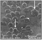
Fig. 25.1
“Quatrefoil” erythrocytes viewed with optical microscopy (black arrows) (×1,000)

Fig. 25.2
“Quatrefoil” erythrocytes viewed with scanning electron microscopy (white arrows) (×1,500)
With regard to breed, the “quatrefoil” RBC pattern was observed in mixed breeds (n = 40; 25.9 %), Labrador Retrievers (n = 18; 11.8 %), German Shepherds (n = 11; 7.2 %), Golden Retrievers (n = 8; 5.2 %), Poodles, Rottweilers, Setters each (n = 5; 3.2 %), Boxers (n = 4; 2.6 %), and others (n = 58; 37.7 %) (Fisher’s Exact test; p > 0.05). The Chi-squared analysis was not statistically significant with regard to sex (p > 0.05) (n = 81 males, 52.6 % and n = 73 females, 47.4 %). Dogs with “quatrefoil” RBCs were split in three age groups: puppy (0–12 months, n = 27 dogs), adults (13 months–6 years, n = 42 dogs), and old (≥7 years old, n = 85 dogs). Compared with the control population, the Chi-square analysis revealed an increased frequency of “quatrefoil” RBCs in older dogs (p < 0.0001). In dogs with “quatrefoil” RBCs, the following alterations were also observed (prevalence in the control population in brackets): anisocytosis (+1/+3), 70.7 % (61.3 %); polychromasia (±/+2), 35.1 % (25.1 %); echinocytes, 22.7 % (20.1 %); rouleaux formation, 9 % (14.5 %); dacryocytes, 9 % (6.4 %); elliptocytes, 7.8 % (6.1 %); acanthocytes, 3.9 % (3.9 %); schistocytes, 3.2 % (4.5 %); eccentrocytes, 2.3 % (1.2 %); keratocytes, 2.3 % (2.1 %); and target cells and spherocytes, 1.3 % (0.7 %), Howell–Jolly bodies, 19.4 % (11.6 %). None of these changes were significantly associated (ANOVA test) with the appearance of “quatrefoil” RBCs.
The ANOVA test performed on CBCs, biochemical profiles and serum protein electrophoresis parameters revealed that leukocyte counts (absolute) and neutrophil counts (absolute and percentage) were significantly (p < 0.0001) lower in “quatrefoil” RBC populations than in the control population.
25.4 Discussion
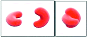
This study is the first to describe the prevalence of these various erythrocyte alterations. The occurrence of “quatrefoil” RBCs is quite rare, about 4 % (154/3,958 dogs). They have been observed more frequently in older dogs, with no differences between breed or sex. These dogs also exhibited more RBC changes (i.e., higher grade of anisocytosis and polychromasia), even though they were not statistically significant (p > 0.05). Compared with the control population, “quatrefoil” RBCs were more frequently found in dogs with lower leukocyte and neutrophil counts. One hypothesis that explains the formation of “quatrefoil” RBCs describes them as EDTA-induced artifacts. Another hypothesis is that dacryocytes overlap with other erythrocytes at the feathered edge of the blood smear. Upon further observation, this hypothesis was rejected because “quatrefoil” RBCs were found with higher frequency in the area between the body of the smear and the feathered edge. In addition, intermediate forms often exhibited round cell characteristics, which were different from dacryocytes. Rouleaux formation was less frequent in dogs with “quatrefoil” RBCs, and thus does not explain the cause of this cell pattern. SEM examination of cell morphology changes confirmed the presence of the “quatrefoil” pattern in blood smears, which disproves the hypothesis of an optical illusion or accidental finding. Furthermore, the SEM analysis provided a basis for a 3-D model. The “quatrefoil” erythrocytes, examined with higher magnification compared to the optical microscopy, displayed an “embraced” cell formation (Fig. 25.3). This pattern is different from the “coin-pile” formation seen in rouleaux formation or the chaotic arrangement seen with agglutinated RBCs.


