Burns
Basic Information 
Clinical Presentation
Disease Forms/Subtypes
• Burns are classified by the depth of the injury:
• First-degree burns involve only the most superficial layers of the epidermis. These burns are painful and are characterized by erythema, edema, and desquamation of the superficial layers of the skin. The germinal layer of the epidermis is spared, and these burns heal without complication.
 Superficial second-degree burns involve the stratum corneum, stratum granulosum, and a few cells of the basal layer. Tactile and pain receptors remain intact. Because the basal layers remain relatively uninjured, superficial second-degree burns heal rapidly with minimal scarring, within 14 to 17 days.
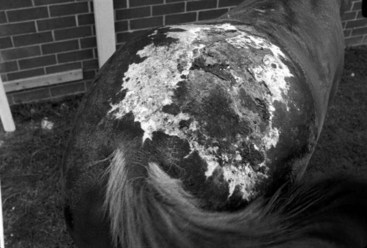
• Third-degree burns are characterized by loss of the epidermal and dermal components, including the adnexa (Figure 1). The wounds range in color from white to black. There is fluid loss and a marked cellular response at the margins and deeper tissue, eschar formation, lack of pain, shock, wound infection, and possible bacteremia and septicemia. Healing is by contraction and epithelialization from the wound margins or acceptance of an autograft. These burns are frequently complicated by infection.
Physical Exam Findings
• Because heat is slow to dissipate from burn wounds, it is often difficult to accurately evaluate the amount of tissue damage in the early phase of injury. Whereas the extent of the burn depends on the size of the area exposed, the severity relates to the maximum temperature the tissue attains and the duration of overheating. This explains why skin injury often extends beyond the original burn.
• Burns are most commonly seen on the back and face.
• Increases in heart and respiratory rates are present in association with abnormal discoloration of mucous membranes.
< div class='tao-gold-member'>
Only gold members can continue reading. Log In or Register to continue
Stay updated, free articles. Join our Telegram channel

Full access? Get Clinical Tree


