23 Blindness
INTRODUCTION
CLINICAL EXAMINATION
Gait, posture, hopping, proprioception and spinal reflexes were normal. No spinal pain was detected.
NEUROANATOMICAL DIAGNOSIS
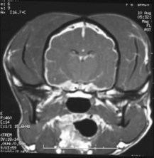
The lesion was localized to the right CNN II, III, IV and VI, and the sympathetic nerve supply to the right eye (Horner’s syndrome) and to the left CNN II and III. The lesion was thought to lie in or near the cavernous sinus, or optic and orbital canals (see page 42).