66 Bilateral atrophy of the masticatory muscles INTRODUCTION Atrophy of the masticatory muscles (MM) allows the skull shape to be more obvious and owners often report a ‘lump’ which is a newly palpable or visible occipital protuberance. Loss of MM leads to enophthalmos. Depending on the cause of the MM atrophy, a dropped jaw or trismus may be seen. SIGNALMENT An 8-year-old male golden retriever dog. CASE PRESENTING SIGNS Generalized muscle loss, especially on the head. CASE HISTORY Diarrhoea, fever and weight loss were noted 1 month prior to referral. The diarrhoea stopped but inappetance, lethargy, fever and weight loss continued. Muscle loss was generalized but particularly prominent on the head which prompted referral. Blood results were abnormal. They showed a normocytic normochromic non-regenerative anaemia, thrombocytopenia, neutropenia, relative lymphocytosis with pleomorphic lymphocytes and occasional atypical large lymphocytes. Alb 23 g/l, ALKP 306 µ/l, ALT 53 µ/l, AST 130 µ/l, CK 259 µ/l, pre-prandial bile acids 25. Prednisolone 25 mg BID was then given for 2 weeks as the referring vet was concerned about MMM despite a low 2M antibody of <1 : 100 (>1 : 100 is consistent with a diagnosis of MMM). The muscle loss did not improve and the dog was referred for EMG. CLINICAL EXAMINATION 
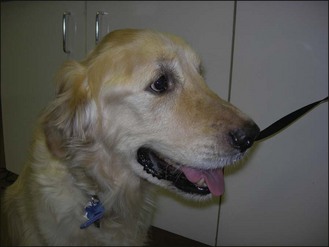
Clinical examination revealed a mentally depressed dog with petechia of the conjunctiva and retina bilaterally. Mucous membranes were pale. The temperature was 39.7°C. There was a generalized muscle loss, including masticatory muscles bilaterally (Fig. 66.1). Jaw tone was good. The dog had normal facial muscle function. Sensation to the face was present. No neurological deficits were found. Figure 66.1 Profound atrophy of the masticatory muscles, pale gums and sclera. Only gold members can continue reading. Log In or Register to continue You may also needMeningitis of large breed dogsComplicated keratoconjunctivitis siccaIdiopathic epilepsySkull tumourReptiles – an introductionHypertensive retinopathyMuscle tone – an introductionHydrocephalus Share this:Click to share on Twitter (Opens in new window)Click to share on Facebook (Opens in new window)Click to share on Google+ (Opens in new window) Related Tags: Saunders Solutions in Veterinary Practice Small Animal Neurology Sep 3, 2016 | Posted by admin in SMALL ANIMAL | Comments Off on Bilateral atrophy of the masticatory muscles