53 AVIAN AND REPTILIAN CYTOPATHOLOGY
1 What are some important principles to understand about the use of cytology in avian and reptilian medicine?
4 List the types of specimens, abnormalities, and lesions that can be sampled for cytologic examination.
6 What are the initial steps involved in evaluation of a cytologic sample?
7 What is neoplasia, and how is it identified in cytologic samples?
Neoplasia generally is identified by the presence and often the predominance of tissue cells with mild to marked nuclear and cellular pleomorphism (Box 53-1). The more pronounced the magnitude of pleomorphism, the greater is the likelihood of neoplasia and malignancy. Inflammation may be present, particularly if the neoplasm is ulcerated or ruptured. A common dilemma in some cytologic samples is how to differentiate inflammation secondary to neoplasia from tissue dysplasia secondary to inflammation.
Box 53-1 Cytologic Criteria for Malignancy
Nuclear Criteria*
Numerous types of neoplasms in all organ systems have been described in reptiles and birds.
8 List the different tumor types and describe general guidelines on their differentiation.
9 What is tissue dysplasia, and how is it identified in cytologic samples?
Tissue dysplasia may be identified in cytologic samples in association with variable amounts of inflammation and is cytologically characterized by minimal to moderate nuclear and cytoplasmic pleomorphism (see Box 53-1). The more intense the inflammatory process, the more pronounced may be the dysplastic changes.
10 How does one differentiate between neoplasia with secondary inflammation and inflammation with secondary tissue dysplasia?
13 What is the significance of heterophilic inflammation?
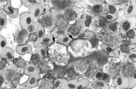
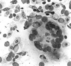
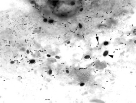
Heterophils predominate (>80% of inflammatory cells) in heterophilic inflammation and indicate acute or recent inflammation. They begin to accumulate in tissues within 3 hours of the inflammatory insult. Heterophils are frequently seen in lesions caused by many types of bacteria (with the notable exception of Mycobacterium). Heterophil degeneration may be detected if bacteria (primary or secondary infection) are part of the inflammatory process (Figure 53-1).
14 What is heterophil degeneration?
Heterophil degeneration is characterized by multiple nuclear and cytoplasmic changes that are unrelated to heterophil toxicity. Degenerate heterophil changes are caused by released bacterial toxins at the site of inflammation or infection. These toxins alter the cell and nuclear membrane permeability, primarily causing swelling and vacuolation (Figure 53-2; see also Figure 53-1). Bacteria may be present within heterophils. If degenerate changes are seen, examine the slide carefully for the presence of bacteria within heterophils or in the extracellular space. Degeneration is characterized by the following:
Karyorrhexis and karyolysis are generally seen with severe heterophil degeneration.
15 What is the significance of mixed inflammation?
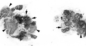
Mixed inflammation refers to an inflammatory process containing a mixture of heterophils (at least 50% of leukocytes), macrophages, lymphocytes, plasma cells, and sometimes, giant multinucleated cells (Figure 53-3). Mixed inflammation is often seen in reptiles and birds. In reptiles, as in birds, mixed inflammation does not necessarily indicate chronicity, since lymphocytes and macrophages may migrate to tissues within a few hours of an inflammatory insult, and multinucleated giant cells form within hours to a few days.
16 What is the significance of a predominantly macrophagic inflammatory process with or without multinucleated giant cells?
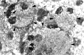
The presence of macrophages and multinucleated giant cells does not necessarily indicate a chronic (several weeks to months) inflammatory process. Conditions in reptiles often associated with a predominantly macrophagic inflammatory process with or without multinucleated giant cells (granulomatous inflammation) include Mycobacterium infection (Figure 53-4), fungal infection, and foreign bodies; xanthomatosis occurs in birds. In contrast to mammals, this type of inflammation may also be seen with many inflammatory conditions, such as bacterial abscesses (birds, reptiles), histomoniasis (turkeys), and chlamydophilosis (birds).
17 When is the presence of bacteria expected, and when is it an abnormal finding?
18 How is cytology used in the diagnosis of bacterial infections?
The main advantage of cytology is that, if present in sufficient numbers, bacteria may be quickly and easily visualized (see Figures 53-1, 53-2, and 53-4). A primarily heterophilic or a mixed inflammatory process (with neutrophil predominance) is seen with most bacterial infections, and heterophil degeneration may be an important feature. The inflammatory reaction to mycobacteria consists of macrophages with or without multinucleated giant cells. Mycobacteria do not stain with Romanowsky-type stains and are seen as negative-staining rod-shaped structures within macrophages and giant cells and in the extracellular space (see Figure 53-4). Acid-fast stains are necessary to visualize the organisms.
19 When is the presence of a fungus expected, and when is it an abnormal finding?
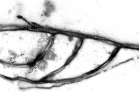
Greater numbers of yeasts, with or without budding, in the upper alimentary tract, feces, and cloaca are an important finding and indicate yeast overgrowth (Figure 53-5). The presence of hyphae associated with yeasts, with or without budding, in the same anatomic locations mentioned above suggests severe infection with probable mucosal damage and invasion.
20 How is cytology used in the diagnosis of fungal infections?
22 How is cytology used in the diagnosis of protozoal infections?
24 How is cytology used in the diagnosis of viral infections in birds and reptiles?
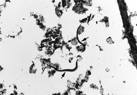
25 How is cytology used in the diagnosis of helminth infections in birds and reptiles?
26 List some diseases of the avian integument for which cytology may be useful in the diagnosis (Box 53-2).
28 What neoplasms of the avian integument can be diagnosed cytologically?
29 What bacterial infections of the avian integument can be diagnosed cytologically?
Mycobacterium spp. do not stain with Romanowsky-type stains (see Figure 53-4). They appear as negative-staining structures within macrophages and multinucleated giant cells. Miscellaneous types of bacteria can also be diagnosed cytologically.
30 What viral infections of the avian integument can be diagnosed cytologically?
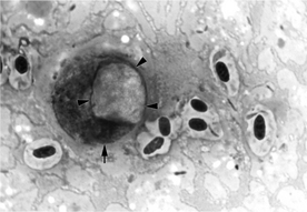
Poxvirus produces intracytoplasmic inclusions that distend infected squamous cell and displace the nucleus paracentrally (Figure 53-9). Poxviral lesions consist of small, crusty areas to discrete masses and are most often seen in the oral cavity and the unfeathered skin. A mixed inflammatory process may be seen.
36 List some diseases of the alimentary tract in birds for which cytology may be useful in the diagnosis (Box 53-3).
37 Can bacterial overgrowth be diagnosed cytologically in birds?
38 What are the cytologic characteristics of intestinal mycobacteriosis in birds?
Romanowsky-stained impression smears and scrapings of intestinal mucosa will reveal macrophages and giant cells distended with numerous rod-shaped, negative-staining organisms in avian mycobacteriosis. Variable numbers of columnar intestinal epithelial cells may be present. Acid-fast stains are necessary to visualize the organisms (see Figure 53-4).
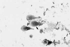
40 Describe the cytologic characteristics of avian trichomoniasis.
Stay updated, free articles. Join our Telegram channel

Full access? Get Clinical Tree


