Chapter 142 Auscultation and Physical Diagnosis
Physical examination follows the obtaining of a complete history and the consideration of the species, age, breed, and sex (see Chapter 1). Diagnosis, or at the very least a number of plausible diagnoses, may be made in many instances by history and signalment; however, following a thorough physical examination, a single, “most likely” diagnosis can be made with reasonable assurance in most patients with cardiac problems.
INSPECTION
Condition, Attitude, and Posture
• Body condition of the patient is classified according to the degree of fat.
• Normal overweight animals usually are not ill from heart failure but may manifest signs caused by pulmonary disease (e.g., chronic obstructive lung disease, pulmonary fibrosis).
• Posture of animals with heart failure is often:
• Standing—reluctant to lay down—with thoracic limbs abducted and neck extended to ease ventilation.
• Swayback with the tail between the legs because of muscular weakness or occasionally caused by digitalis toxicity (from overdosage or impaired renal excretion related to dehydration or primary renal disease). Swayback can also result from electrolyte imbalance—in particularhypokalemia—caused by prolonged or excessive use of diuretics without consumption of sufficient electrolytes.
Mucous Membrane Color
• Normal mucous membranes have a deeply saturated pink color. Gentle pressure on the gums will lead to blanching (whiteness) of the membranes. The capillary refill time needed to regain normal coloration is about 2 seconds or less.
• Delayed refill time generally indicates peripheral vasoconstriction, often as a compensatory response to reduced arterial blood pressure.
• Bright red membranes associated with a very short refill time may indicate peripheral vasodilation, as might occur with septic shock or during treatment with potent arterial vasodilator drugs.
• Mucous membranes are pale in some patients. Pallor indicates anemia or low cardiac output with reflex vasoconstriction (e.g., low output heart failure, severe aortic stenosis, cardiac tamponade), but without pulmonary congestion that often causes cyanosis.
Cyanosis
• Methemoglobinemia (congenital or acquired from drug toxicity) may also alter the color of the mucous membranes.
• Cyanosis to the mucous membranes may be observed in late stages of congestive heart failure or in puppies or young dogs with tetralogy of Fallot or other congenital heart defects with right-to-left shunts. (see Chapter 154)
Abdominal Distention and Edema
• Abdominal distention may be a result of gas in the gastrointestinal (GI) tract from aerophagia associated with chronic pulmonary disease, respiratory failure, primary GI disease, obesity, abdominal wall weakness (e.g., Cushing’s disease) or fluid accumulation.
• Ascites and enlargement of abdominal organs (especially the liver) may indicate systemic venous hypertension or passive congestion due to right-sided heart failure or pericardial disease.
• Pitting edema of the limbs, brisket, and prepuce can be caused by severe right-sided heart failure.
Pattern of Ventilation
• Rate of breaths per minute should be between 12 and 20 for dogs and only slightly higher for cats, but may increase from excitement, fever, anxiety, warm environment, chronic respiratory disease, respiratory failure, left-sided heart failure, or pulmonary injury.
• Depth of ventilation is difficult to quantify, but hyperpnea (increased depth) is often a sign of blood gas derangement (e.g., response to metabolic acidosis). It is not often observed in heart disease except with severe pulmonary edema. In upper airway obstruction both the inspiratory and expiratory phases of ventilation may become slower and exaggerated. In pleural diseases, the depth is generally shallow and the rate rapid. An exaggerated expiratory phase is typical of bronchial obstruction.
• Dyspnea (labored ventilation or respiratory distress) may occur as increased rate and/or depth of ventilation or merely as increased effort. With dyspnea one observes:
• Pulmonary retraction is an indention of the thoracic wall caused by fatigue of muscles of ventilation. Regions of the thoracic wall affected must generate greatest tension during ventilation and do not have assistance from thoracic limbs. Pulmonary retraction produces an “hourglass” configuration to the thorax (most evident when viewing the dorsoventral radiograph).
Cough
Cough is a sign of both heart and lung disease. It may be characterized as follows:
• A hacking, honking, brassy cough indicates disease of the large airways, such as tracheobronchial collapse, compression of the left mainstem bronchus due to mitral regurgitation or generalized cardiomegaly, tracheobronchitis, or bronchopulmonary parasitism. This type of cough is seldom due to injury of the parenchyma of the lung and therefore is uncommon in pulmonary edema or pneumonia.
• Subtle or “half-hearted” coughing may be caused by pulmonary edema, pneumonia, or diaphragmatic hernia.
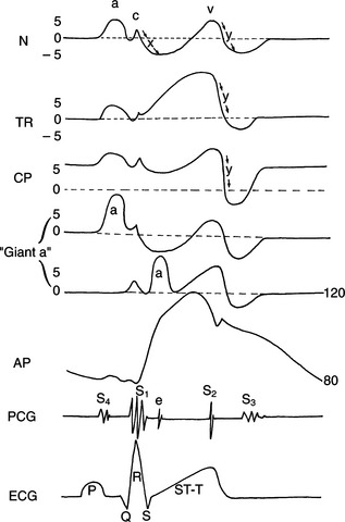
Jugular Vein Evaluation
• The jugular vein normally collapses totally when the long axis of the head is tilted up at an angle of 45 degrees with the horizontal.
• The jugular vein also normally collapses during inspiration and immediately after the second heart sound.
• If the jugular vein does not collapse, the venous pressure may be elevated owing to heart failure, chronic obstructive pulmonary failure (COPD)/pulmonary fibrosis (PF), or ventricular filling restriction due to pericardial disease.
• A firm, distended jugular vein that collapses briefly immediately after the second heart sound is consistent with cardiac tamponade from pericardial disease. (see Chapter 151) Causes in dogs include hemorrhage into the pericardial sac due to a bleeding neoplasm or rupture of the left atrium with severe mitral regurgitation. Constrictive pericarditis and chronic respiratory disease also can cause similar jugular venous changes.
Vigorous Pulsation of the Jugular Vein
• Jugular venous pulses (“cannon a waves”) occurring at rates exceeding 120/min with a very slow ventricular rate slow (usually below 60/min) indicates third-degree (complete) or high-grade second-degree atrioventricular block.
• If cannon a-waves occur just before the first heart sound it may rarely indicate tricuspid stenosis or possibly stiffness of the ventricles (e.g., pulmonic stenosis).
Hepatojugular Reflux
• Pushing on the abdomen of a standing dog displaces the liver dorsad and “milks” blood from the liver to the right side of the heart, increasing systemic venous return.
• If the right ventricle cannot pump the extra blood through the lungs, or if right ventricular filling is restricted owing to pericardial tamponade or constriction, the extra venous return “wells up” in the atrium and systemic veins. This situation produces added jugular venous distention or a positive hepatojugular reflex.