47 Ataxia
Spinal ataxia
INITIAL PRESENTATION
In spinal cord disease, the ascending proprioceptive tracts may be affected before the descending motor tracts resulting in ataxia as the presenting sign. Damage to the ascending spinocerebellar tracts in spinal cord lesions results in cerebellar-like dysmetria. Spinal cord lesions will cause ataxia of the limbs caudal to the lesion.
INTRODUCTION
Coordination relies on balance, position sense and the ordering of received sensory information into smooth movement. Failure of coordination, from any of the components, results in ataxia a term used to describe irregular, unpredictable voluntary movement.
Causes of ataxia
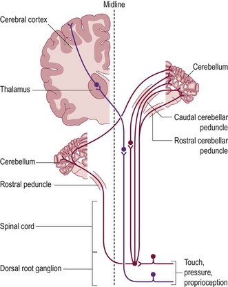
Position sense is referred to as proprioception and is achieved through muscle, tendon and joint receptors, associated sensory nerves, and ascending tracts within the spinal cord (Fig. 47.1).
These tracts project to the ipsilateral cerebellum (unconscious proprioception) and to the contralateral cerebral cortex (conscious proprioception). This constitutes ‘general proprioception’.
When this system fails, the gait becomes ataxic and limb position alters. This is most noticeable with spinal cord and brainstem lesions with deficits ipsilateral to the lesion. Cerebral lesions are subtle and tend not to affect the gait but can reduce the contralateral placing response. Sensory neuropathies are rare. Ataxia and anaesthesia of the limbs with self-mutilation are the two chief clinical presentations of sensory deficits. Lesions of muscle, tendons, and joints present as lameness not ataxia.
Presenting signs of abnormal proprioception
Paresis may be responsible for the inability to support the weight and walk normally, resulting in stumbling, tripping or collapse. Supporting the animal’s body weight while testing placing or the knuckling responses should distinguish a proprioceptive deficit from paresis.
Owners commonly describe their pet as ‘drunk, wobbly, clumsy or unsteady’.
CASE HISTORY
The dog suddenly started yelping when touched and would not jump into the car 3 weeks prior to referral. He was normally an active dog and spent the working day around horse stables. His condition did not improve with self-imposed rest and he remained lethargic and inappetant. When walking, the dog kept his head lowered and his back arched.
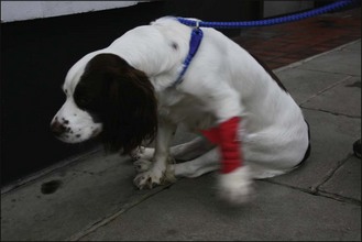
Hindlimb ataxia began 6 days before presentation and slowly worsened. The dog was unable to walk more than a few steps before the hindlimbs crossed and tripped each other (Fig. 47.2).
CLINICAL EXAMINATION
The dog was quiet but mentally responsive. He was unable to stand without assistance. The back was arched, and pain was elicited by palpation of the lumbar spine. Hindlimb paresis and ataxia were present when the body weight was supported with a sling. The right hind knuckled over more than the left. Spinal reflexes were intact. The forelimbs were normal. No neck pain was found.
DIFFERENTIAL DIAGNOSIS
IVDD, external trauma, meningitis or discospondylitis could all give rise to a sudden onset of pain. A migrating grass seed within spinal muscles with cellulitis would cause back pain but extension to the spinal canal would be unusual. No vertebral fractures could be seen on the radiographs taken by the local veterinarian.

Stay updated, free articles. Join our Telegram channel

Full access? Get Clinical Tree

